
When you think 3D you probably imagine the cinema and popcorn, or that fancy TV you’ve just blown the kids’ university fees on. What you probably don’t think - unless you’re a particular breed of palaeontologist – is molluscs. And certainly not printing them out in 3D.
But this practice, strange as it seems, is becoming increasingly common, with some startling applications.
A recent study by University of Texas researcher Jakob Vinther and colleagues is a wonderful example of the high-tech tools many modern palaeontologists use to understand fossils.
This study, on a primitive group of molluscs, employs a number of different techniques – traditional observation, high resolution CT scanning, computer reconstruction and DNA-based dating methods – to better understand the evolutionary relationships and biology of this fossil group. And, yes, some 3D printing.
The animals
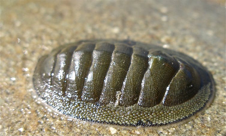
Vinther and colleagues describe a new species in an extinct group called the multiplacophorans. These are molluscs (a larger group that includes mussels, squid and snails) which had a shell on their back, split into 17 plates.
These plates sat on the soft parts of the animal – a thick, leathery mantle, which had many smaller hard plates or spines embedded around the edge. The fossil used by Vinther and team is a 390 million year-old specimen from Ohio.
The researchers used the anatomy of the creature, combined with DNA-based dating, to suggest the fossil – and the group to which it belongs – is most closely related to a living group called the chitons (or polyplacophorans) but is not a true member of that group.
From this they surmise some of the similarities between the groups must have evolved separately – they are an example of convergent evolution, when similar selective pressures result in animals independently evolving similar traits.
One of the tools the authors used to understand these creatures – and reach these results – is X-ray micro-tomography (µCT) – a high resolution form of CT scanning.
The technique
µCT is a powerful and increasingly mainstream tool employed by palaeontologists to study 3D fossils. While the majority of fossils aren’t preserved in 3D, those that are can often prove difficult to study – bits of the animal can remain buried in the rock.
µCT is a non-destructive, X-ray based technique that helps overcome such limitations. By taking a large number of X-rays (or projections) of a specimen as it rotates, µCT can create a series of slices, or a 3D volume, which maps the interactions between different materials and X-rays.
This allows a digital 3D model to be created.
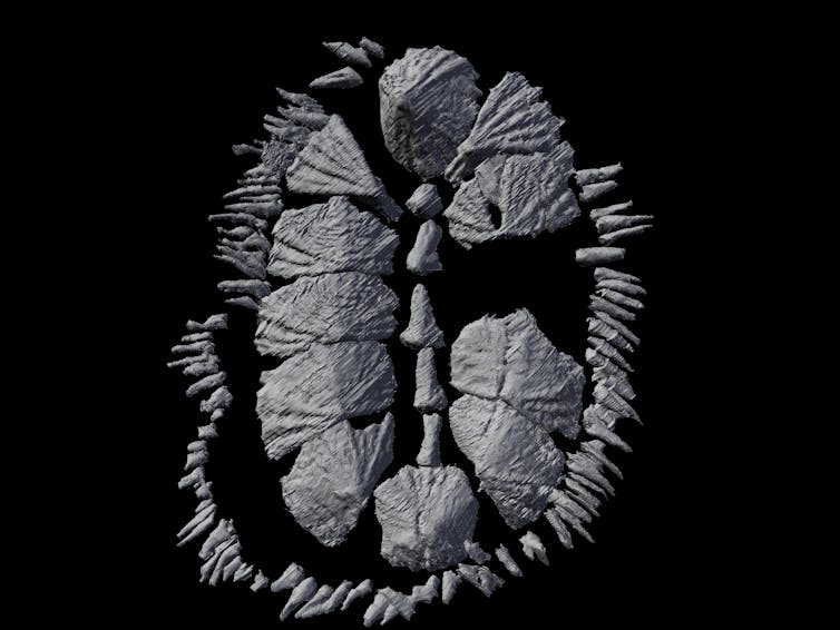
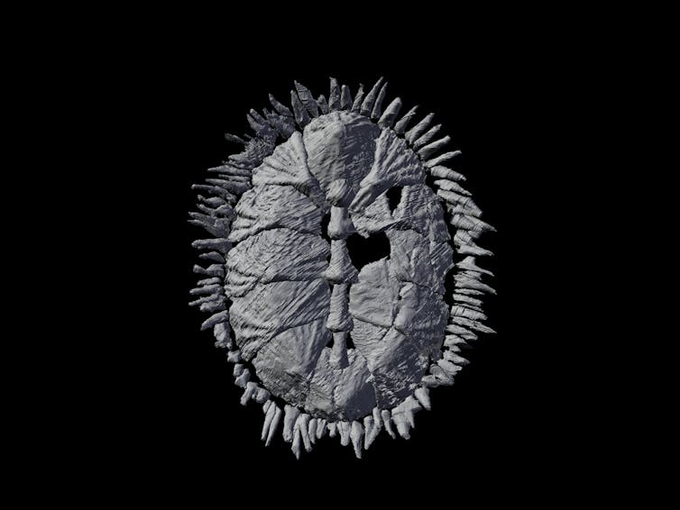
The mollusc fossil studied by Vinther and colleagues wasn’t perfectly formed. When the creature died and rotted its plates separated, leaving a disjointed fossil. After creating a 3D digital model (including portions of the plates which were buried in rock) they used software to reassemble the disarticulated fossil.
And that’s not all …
The reconstruction
Vinther and colleagues made further moves to understand the long-dead creature by creating a physical reconstruction. First they used a 3D printer to create a physical representation of the digital model.
This piece of kit is a machine that takes the digital files that record the fossil’s 3D anatomy and uses them to build a solid model of the organism.
This technique comes in many flavours, but the type palaeontologists use usually involves plastics or resins, either liquid or powder in form. The material is then fused to create a solid object in the correct shape, most commonly using lasers.
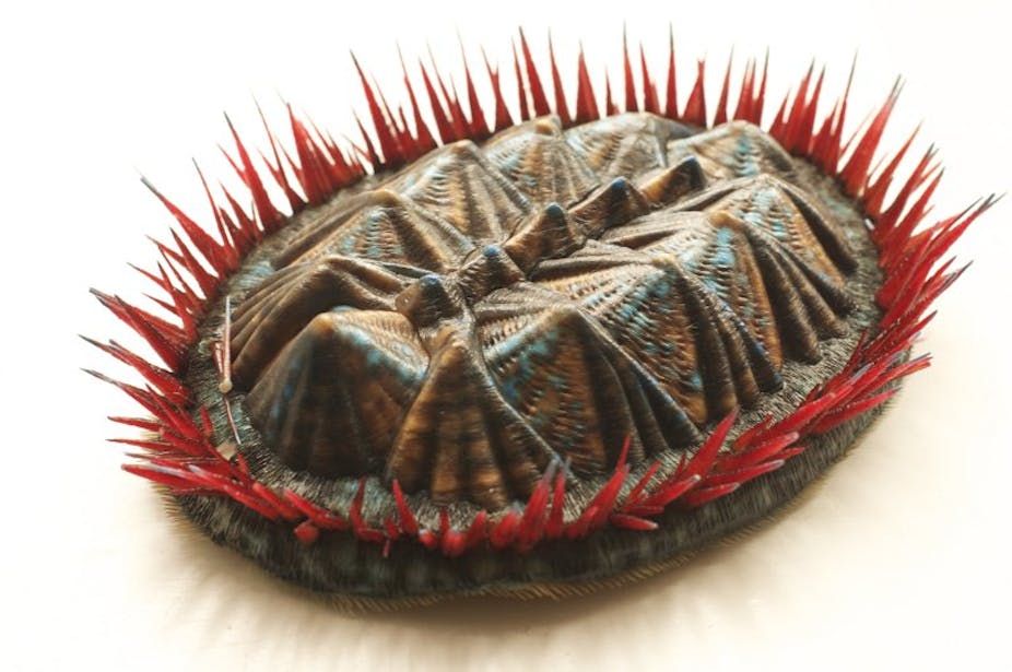
The end product is a physical 3D model – in this case a reassembled multiplacophoran, twelve-times larger than the original. The final stage of this process was to create a realistic recreation of the animal in life, which was done by hand, with clay, plastic and lots of paint.
The future
In the last decade palaeontologists have been applying X-ray techniques, and the other methods mentioned above, to a wide range of creatures, from the tiny earliest preserved living animals to more recent, and far larger, dinosaurs.
Clearly, using such techniques can help us better understand fossils. Through resolving their anatomy in full, helping us recover body parts from the rock and sometimes even allowing us to see their internal organs, the methods now being mastered are giving us a clearer picture of extinct animals’ biology.
As the study by Vinthers and colleagues admirably shows, we can also gain a clearer picture of those creatures’ evolutionary relationships, and we can see long-dead organisms in almost as much detail as if they were alive today.
Now, surely that beats a trip to the cinema …