
The human brain is the most complex arrangement of matter in the known universe. Through our five senses it “digests” vast amounts of information that allows us to see, hear, taste, touch and balance. It commands our muscles, it learns, remembers, hungers, loves and hates.
Understanding how the brain works is a major research challenge; thousands of scientists are studying it in the expectation that through greater understanding we can eventually overcome many tragic diseases and injuries.
What goes wrong during stroke or in dementia? What are the causes and genetics of brain disease, age-related hearing loss, motor neuron disease? What treatments will improve them and will psychological interventions help? Then there are the those who want to explore the brain, to find out how it ticks and how, for example, we see and read.
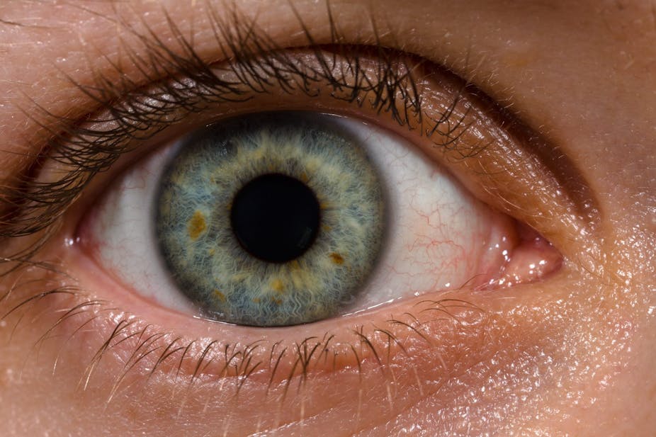
The eye is the only part of the brain that can be seen directly – this happens when the optician uses an ophthalmoscope and shines a bright light into your eye as part of an eye examination. It shows the innermost layer of the eye (the retina), and the nerve carrying visual messages from the retina to the brain (along the optic nerve) are visible in the back of the eye.
In many neurological diseases, such as multiple sclerosis or stroke, we can see changes in the optic nerve that provide a direct diagnosis. And if pressure in the brain increases, perhaps due to a brain tumour, we can see this as a swelling of the optic nerve. So changes in the back of the eye can be used in the diagnosis of high blood pressure, diabetes, glaucoma, age-related macular degeneration or genetic diseases, such as retinal dystrophies.
An immature retina
A new sophisticated method called optical coherence tomography (OCT) allows the retina to be measured with much more detail. We can see all ten retinal layers and scan them in a few seconds at almost microscopic resolution. OCT has greatly enhanced diagnosis and treatment of retinal and optic nerve diseases in adult patients. For example in glaucoma, loss of nerve fibres can be detected, and assist diagnosis and detection of progression.
Until recently it was not possible to examine children with OCT because they could not stay still for long enough. However, hand-held scanners are much more suitable for children. But there is little knowledge about what constitutes a “normal” retina in a child and of course things are also changing as children grow and develop.
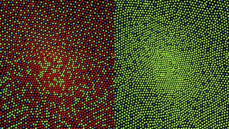
At the University of Leicester Ulverscroft Eye Unit researchers are investigating normal and abnormal development of the retina and optic nerve as an aid to future diagnosis. They found that the retina is immature at birth. For example, the photoreceptors, the sensors in the retina which detect light, are very small at birth, but slowly grow and elongate by about 30 times, until the age of six.
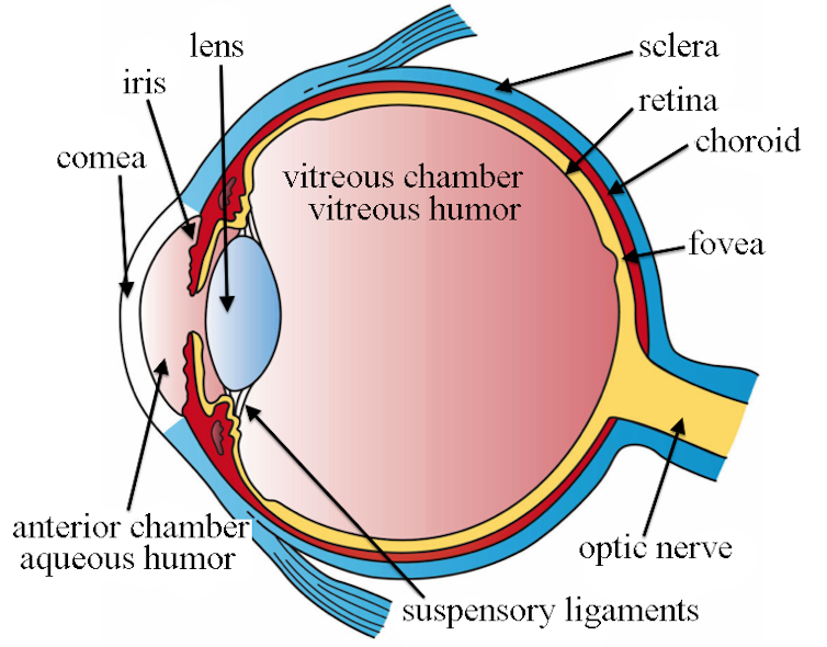
Scientists can also follow the development and formation of the fovea (this is the central region of sharpest vision, made up of closely packed cones) and this shows that retinal and optic nerve development is not completed before young adulthood.
Abnormal photoreceptors
In several eye diseases the normal development of the fovea is slowed or halted. The photoreceptors remain small and the inner retinal layers do not migrate properly. This is called foveal hypoplasia. Disrupted development can occur in genetic diseases, for example in albinism, or in very early born premature babies.
In these children vision is reduced. Often they cannot hold their eye still and develop constant involuntary to-and-fro movements of the eyes, called nystagmus. In retinal dystrophies, the lining of the retinal layers is disturbed. Abnormal foveal development and retinal changes can be diagnosed very early with OCT. Early diagnosis can direct further exams and genetic investigations and help to avoid unnecessary examinations under anaesthesia.
The eye is the only part of the body where nervous tissue and vessels can directly be seen. This allows direct view of changes caused by disease. The direct view of the retina and optic nerve make the eye extremely well suitable for research, for example to observe maturation of nervous disease and changes in neurological disease.