

As a sports and exercise biomechanist who has traditionally worked with professional athletes, it came as something of a surprise when the Mary Rose Trust contacted me back in 2011. The charity asked me to analyse the skeletons of men who drowned aboard the Mary Rose battleship in 1545. But these were no ordinary men, they were professional archers, and so could also be considered elite, or even ultra athletes, trained to go into battle for the then King of England, Henry VIII.
The research involved scanning the bones to produce very precise, virtual replicas. We analysed these replicas, minimising the need to handle bones directly. Our aim was to determine if, by precisely measuring the bones, we could identify which of the remains were the archers. This work – which was later used to put together a reconstruction of one archer’s face from a skull scan – was the very beginning of research which we now hope will unlock the past for scientists and historians all over the world.
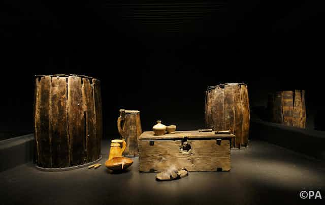
The Mary Rose lay at the bottom of the Solent, the strait that separates the south of England from the Isle of Wight, for nearly 450 years after its sinking – until 1982. Since then some 19,000 artefacts have been recovered from the site of the wreck, ranging from cannons to fiddles, earscoops to hilted swords. Due to the conditions in the Solent, the ship very quickly became covered in silt and mud after it sank. This meant that it, the artefacts and the skeletons of the crew were incredibly well preserved. In fact, the state of preservation was such that some of the longbows recovered from the ship were still usable.
Yet like any item with historical merit, these are still incredibly delicate objects, which need to be handled carefully so as not to damage or contaminate them. They are in much demand for examination, meaning that researchers may struggle to get time with the items in person. However, by creating a virtual 3D database of detailed scans that can be accessed by researchers the world over, we hope that more experts with different areas of knowledge can access the items, and contribute to the analysis of them.
History in 3D
Using 3D scanning and imaging to produce models is not, it has to be said, a new concept. Museums have been using this innovative way of displaying artefacts for some time. Others, like Toby Jones, curator of the Newport medieval ship project, have even used imaging as a tool to accurately reconstruct the dimensions of and preserve whole ships digitally, piece by piece. We are taking this one step further with our Mary Rose work, and making not only a full database of the artefacts, complete with 3D images, but a resource for the scientific community to access and study. Our scans of items and skeletal remains from the ship are being produced to challenge the research community and, in particular, see if a full analysis of the bones – the likes of which has only been achieved with a first hand examination in the past – can be achieved from a digitised archive.
When we examined the archers’ skulls, a laser scanner was used to create exact three-dimensional virtual replicas. For this latest work, we decided to do things differently, instead opting for photogrammetry as the means of digitising the artefacts. Photogrammetry is the use of photography to map and survey objects, here resulting in 3D digital models of each artefact recovered from the Mary Rose. We chose photogrammetry over the previously used laser scanning as this time we were not interested in measuring dimensions of the skulls, but in the visual data. Photogrammetry is ideal for this as the photo-realistic images can be manipulated by the user.
The project’s PhD student Sarah Aldridge took the task on, carefully photographing every skull around 120 times each, using a 39 megapixel camera. Items with higher aspect ratios, or different shapes, required many more photos: around 400 images of the heavily detailed carved wood panel were taken. The photos were then edited and combined using software to create detailed representations of them.
Digital vs. real life
As our work progressed, we asked a group of archaeologists to analyse the scans of real skulls, and virtual skulls made using photogrammetry. Though this study is not yet complete, our initial results are very promising and showed which traits were recognised well and not so well using the photo technique.
Some skull properties, for example, are typically analysed to determine gender or ancestry, and are more tactile, traditionally requiring a close examination. The upper edge of the eye socket is one such feature: the sharper the edge, the more feminine it is considered. By conducting the study in a controlled environment, we were also able to optimise the method for viewing the digitised image. This was achieved by ensuring that the laptops used were correctly calibrated and of sufficiently high resolution to faithfully reproduce the nuanced 3D models.
At present only those working in the field of bone science – osteologists, forensic anthropologists, bone biologists and the like – have access to the research sections that we have published on our website, however, we hope that more will be open to the public in the future. Going forward, the work that we have done on the Mary Rose artefacts could open up a whole new method of scientific analysis, allowing researchers to examine any artefact from anywhere in the world at any time.