

The first cases of AIDS were described over 30 years ago, in the early 1980s. The notion of a new viral disease that killed almost everyone who became infected was terrifying. Since then, HIV infection has become a manageable chronic condition, with a normal life expectancy if one is lucky enough to be able to access treatment. HIV is now the most studied and best understood virus of all.
A virus is the smallest biological entity that can replicate itself, although they are absolutely dependent on their host. In the case of HIV, only humans are infected. Viruses are essentially molecular machines that can reprogramme host cells to make viruses.
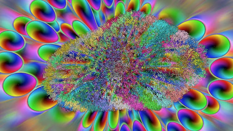
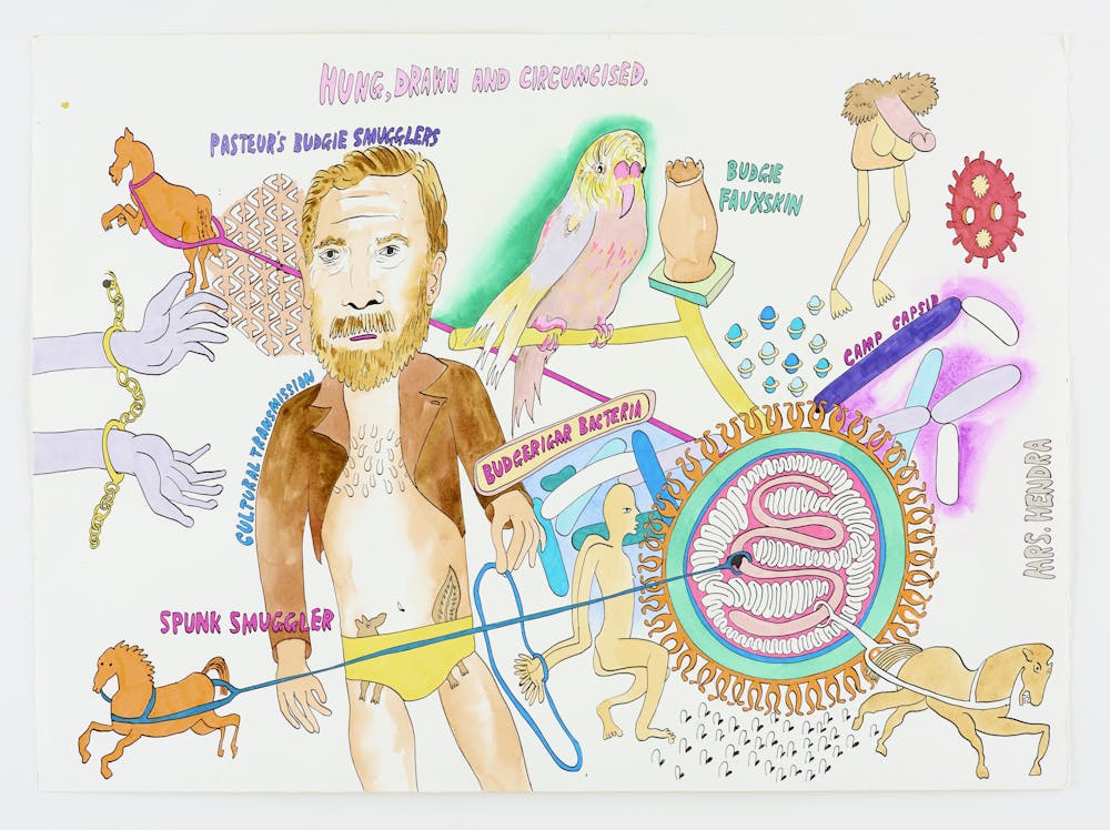
Viruses are much too small to see with the naked eye. Scientists visualise them using powerful microscopes which use beams of electrons instead of light. This fast developing technology is revealing virus structure in ever greater detail, helping us understand how viruses work. But there are other ways of visualising viruses too, as we discovered when we began working with the artist John Walter. He got in touch about some of our microscope images of HIV.
Viruses
All living things are infected with viruses. Viruses typically cause disease in their host. The host, in turn, typically deploys a multi-layered defence system to prevent and control infection. This is our immune system. Viruses and hosts are engaged in an antagonistic relationship that has existed throughout the evolution of life on earth. This is evidenced by remnants of ancient viral infections, which left their DNA in host genomes and now comprise about 8% of human DNA.
The work in our laboratory rests on the broad hypothesis that HIV has been able to infect 80m people, kill 30m, and cause a global pandemic, because of its unique ability to overcome the defences encompassed by our own immune system.
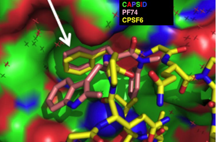
We work on the HIV capsid. The capsid is a container that protects the viral genetic material. But it is not a passive container. It is a molecular machine that regulates synthesis of viral DNA and acts as a GPS by interacting with a series of proteins in the host cell that it infects. Through these sequential interactions, the virus knows where it is, what it has to do and when to do it. We aim to understand how the capsid works, asking how it interprets this information and how we can target capsids with new types of drug.
Capsids
We began working with John when he contacted us to ask us questions about capsids. John had been making art influenced by the HIV epidemic for many years, and his work had begun to consider what he describes as “the crisis of representation in HIV”. John wanted, as he put it, to “look under the bonnet of HIV”.
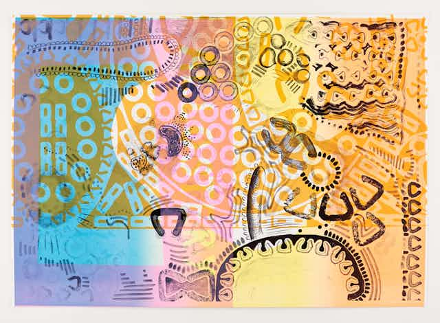
He found out about capsids. As a central part of our work, we make images of capsids coated with their host cellular protein binding partners. John had seen some of these pictures and had even made some beautiful viral capsids out of cardboard. We were struck by how accurate they were.
We realised almost immediately that despite our different perspectives, we were asking the same questions. How does the capsid fit together and how does it work? Somewhat to our surprise, our early conversations quickly turned into a genuine collaboration. John attends our lab meetings, asks questions about our work and inspires new questions and perspectives. John acts as the jester. His questions empower us to think afresh about our observations and their interpretation. John licenses our creativity in a new way, and we hope that we do the same for him.
John’s art is massively influenced by the discussions he has in the lab. But he doesn’t just illustrate our work. Rather, our science provides new material to inspire his work.
Painting science
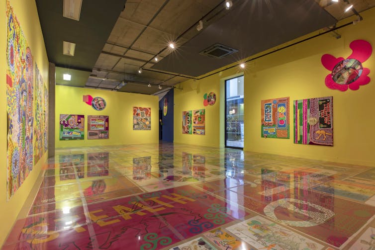
For example, John has produced epic paintings of the inside of a cell as seen by a virus. His “cytoplasm” series is influenced by our discussions of hostile hosts and evasion of defences and cloaking of viruses from innate immune systems. He has also made a film called A Virus Walks Into A Bar that tells the story of how a capsid is assailed by host defence mechanisms but is eventually successful, infecting the cells and drinking at the bar. Some of us are in the film.
His “allostery” series of works, meanwhile, are influenced by our hypothesis that the HIV capsid changes shape each time it touches part of a host cell. Allostery is the word we use to describe shape changes when proteins touch. Critically, it’s not just the touching bits that change, the whole protein can change shape. We propose that this is how the HIV GPS works. It knows where it is depending on what shape its capsid proteins are in.
Science and art are both highly creative disciplines and our work with John has really brought this home to us. It has promoted a real commitment to taking a creative approach to answering our scientific questions. John has given us a new basis for encouraging early career scientists and students to recognise that science is a creative enterprise. He now teaches our undergraduate students and discusses scientific problems with our PhD students and post docs.
Our collaboration with John has been much wider-reaching than we expected. We have encouraged each other to expand the space in which we ask questions and open ourselves to other perspectives. We think, perhaps, that we are inventing a new way to do science, and a new way to do art.