


How do we take aim at things? For example, how are athletes able to accurately throw a javelin, throw a boxing punch or put a ball into the 18th hole?
Many sports involve aiming and rapidly delivering an object, like a ball, that goes out of control once it’s released. In these situations, people must take aim based on initial visual information. They happens in two ways, either relative to themselves (something called egocentric direction) or relative to a visual landmark (allocentric direction).
Egocentric direction is an automatic process. It involves calculating an object’s direction relative to your eyes and keeping track of your eye position when launching the object. Allocentric direction involves seeing and remembering an object relative to some familiar and stable object, like the goalie relative to the goalpost.
In real world conditions, both of these mechanisms are combined in the brain. And ultimately the brain must convert both into egocentric commands to send signals to tell muscles to contract and carry out the action.
There are many examples of this in sports. To score a point, a soccer or football forward on a breakaway must use allocentric information, like where he sees the goal keeper relative to the goal, and the ball relative to his foot. But he must also use egocentric information, like his sense of goal location relative to his eye, and his foot relative to his body. The brain then combines all this information to trigger a kick that sends the ball toward the open part of the goal.
Which parts of the brain?
The egocentric mechanisms that visually guide action are fairly well known by neuroscientists, but relatively little is known about allocentric ones.
It is believed that more dorsal (upper) parts of the visual brain are involved in transforming egocentric vision into muscle commands, while more ventral (lower) parts of the visual brain are allocentric. This thinking is mainly based on studies with brain-damaged patients or ones that involve visual perception. But the specific brain areas that are involved in remembering allocentric visual locations to guide manual actions, like reaching toward an object, haven’t been investigated.
We aimed to do this in a recent study, published in the Journal of Neuroscience. We monitored brain activity in participants using functional magnetic resonance imaging (fMRI) while they glimpsed a briefly presented target, then remembered its location for several seconds, either relative to themselves (egocentric) or to a visual landmark (allocentric), until finally they were cued to aim a reach in the dark.
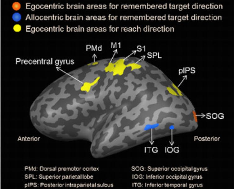
The results showed that partially overlapping but different brain areas were used for both types of visual processing. In particular, when participants were instructed to remember egocentric target locations, a part of the cerebral cortex at the back of the brain, called the superior occipital gyrus, encoded their visual direction. In contrast, the allocentric task caused activity in areas called the inferior occipital gyrus and inferior temporal cortex, to the lower and near the sides of the brain. In both types of task, other areas in parietal and frontal cortex, near the top and front of the brain, coded direction during the final reach response.
Real-world applications
These are scientifically novel results with clear implications. Besides helping to explain what is going on in the brain in demanding situations like sports, there are clinical applications. If the egocentric brain areas are damaged by a stroke, for example, the allocentric areas may still be able to influence action. In these patients, rehabilitation therapies designed to enhance the use of allocentric cues could reinforce recovery and continued function.
Likewise, egocentric function requires constant fine-tuning (through practice) of the brain’s internal sense of vision, eye position, and other body parts. This ability likely degrades in neuro-degenerative disorders and even during normal ageing, so strategies that build on allocentric mechanisms may be helpful for these people too.
So we know what areas of the brain are used when taking aim and how we might use it to help people with certain conditions. But what remains a mystery is how the allocentric parts of the visual system are able to influence parts of the brain that control actions like reaching. In other words, what is going on in the brain at the moment the athlete decides to take a shot at the net? To figure out this puzzle, we plan to go back to the lab to do more experiments.