


Widening our view of the world can mean taking a much closer look at the familiar. And technology from MRI to Scanning Electron Microscopes, which use focused beams to interact with a sample’s surface to produce nano-sized resolution, is allowing scientists and medical researchers to delve into our strange and beautiful world (sometimes aided with a little Photoshop).
Three academics with winning entries in this year’s Wellcome Image Awards tell us how they got their image.
Kevin Mackenzie, Microscopy Manager, Institute of Medical Sciences, University of Aberdeen
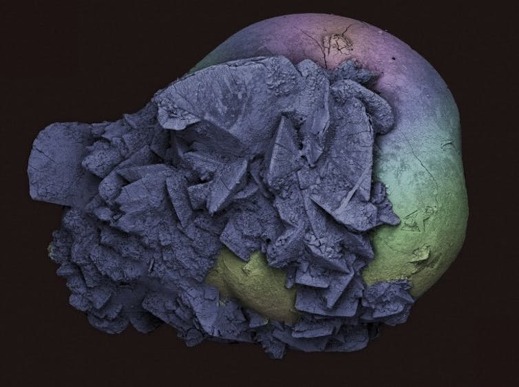
I’m always on the lookout for new and interesting specimens to image and when I had kidney stones a few years ago I managed to collect one. I decided to image in the light microscope, Micro CT and also under the Scanning Electron Microscope (SEM). The resulting image was taken using a Zeiss MA10 SEM and false coloured using Adobe Photoshop. The size of the stone is 2mm across (which is quite small for a kidney stone).
Kidney stones form when salts, minerals and chemicals in the urine (for example calcium oxalate and uric acid) clump together and solidify. Small kidney stones are often passed naturally, but larger stones sometimes get stuck in the kidney or in the tubes that carry urine out of the body.
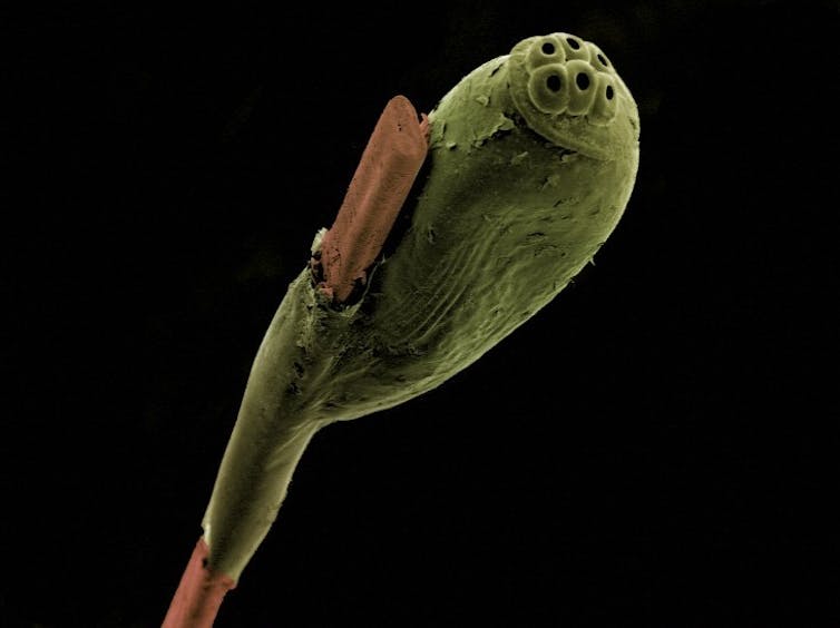
I was very happy to have another two images selected for the awards, one was a Scanning Electron Micrograph of a single head louse egg attached to a human hair, and the other was a Micro CT scan of a medieval (so over 1000 years old) jawbone.
Zeynep Saygin, Postdoctoral Researcher in the Department of Brain and Cognitive Sciences at MIT
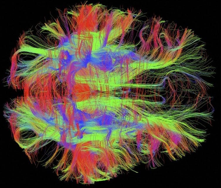
My image depicts the nerve fibres, or wiring, of the healthy human brain. Brain cells communicate with each other through these fibres and we can visualise them in every individual using a specialised MRI scan. The colours represent the direction of the fibres: blue for those that travel up and down; green for front to back; and red for left to right.
I am continually astounded by the number and complexity of these nerve fibres; what information do these connections carry and how do they orchestrate complex mental processes? My research combines these images of connectivity with neural response patterns in order to understand the complex circuitry of the brain, and how it ultimately shapes who we are.
Sergio Bertazzo, Junior Research Fellow, Imperial College London
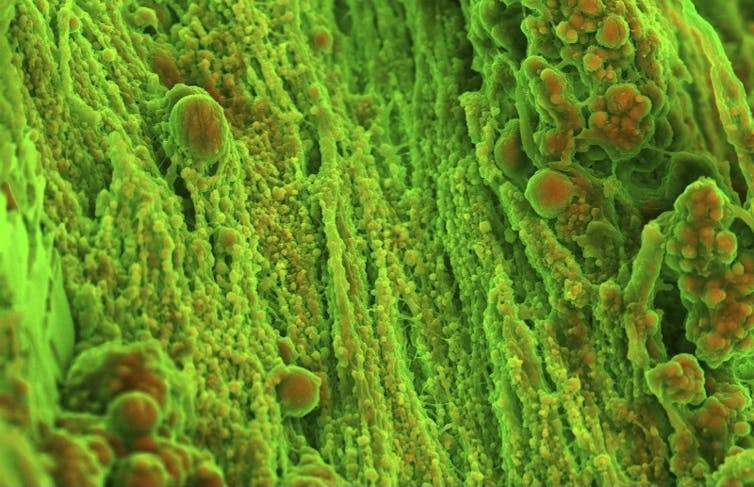
This is a density-dependent colour scanning electron micrograph of the surface of human heart (aortic valve) tissue. The spherical particles show calcification. The orange colour identifies denser material (calcified material composed of calcium phosphate), while structures that appear in green are less dense (corresponding to the organic component of the tissue).
The discovery of calcified particles shows that calcification in the cardiovascular system is more complex than being just a regular process of bone formation.
Best of the rest
Anders Persson, Director of the Centre for Medical Image Science and Visualisation at Linköping University, Sweden
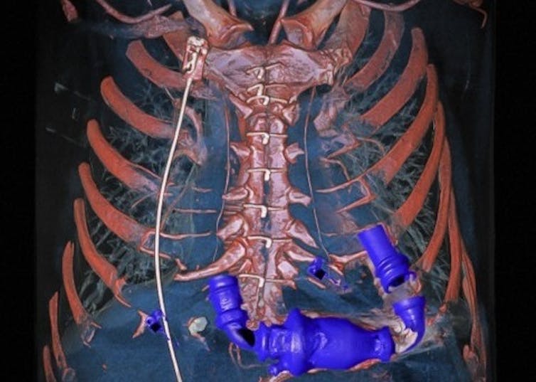
The overall winner, this image was created from a new type of scan called dual energy computed tomography (DECT) angiography. Unlike CT scanning, DECT uses two sources of X-rays at different energies. These are then digitally reconstructed in 3D and can be rotated, sliced or magnified.
Khuloud T Al-Jamal, Senior Lecturer in Nanomedicine, and Izzat Suffian, PhD, King’s College London
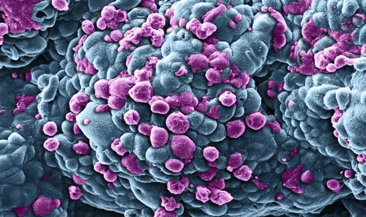
Scanning Electron Micrograph of a cluster of breast cancer cells (in blue) treated with nanometre-sized particles that carry the anti-cancer drug doxorubicin. This causes some of the cells (in purple) to die through a process known as programmed cell death, where cells effectively commit suicide in a controlled, predictable way. Turning this on in cancer cells can reduce a tumour’s size.
Annie Cavanagh and David McCarthy, Microscopists
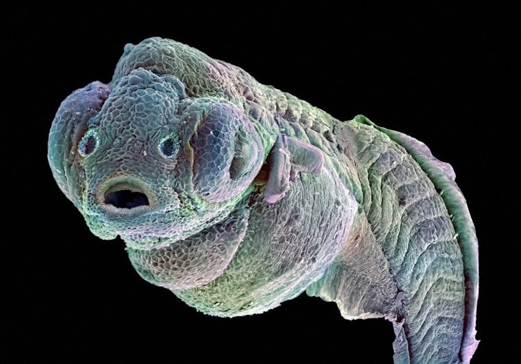
Scanning electron micrograph of a four-day-old zebrafish embryo. To capture this image, the zebrafish was physically attached to a stub (specimen holder) by its tail and tilted to 65 degrees. As zebrafish embryos are approximately 1cm in length, making the whole embryo too big to be captured in a single image, three separate images had to be taken along its length and then stitched together digitally. Colour was then added to the black-and-white image.
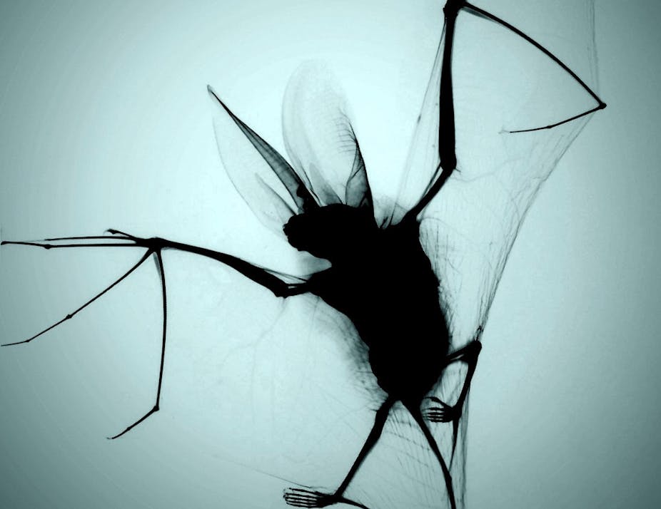
Chris Thorn, Medical Artist
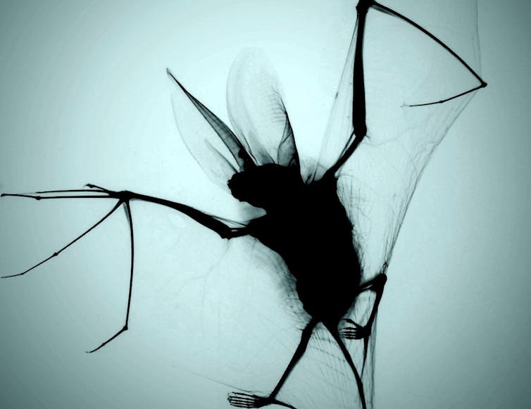
X-ray projection of a brown long-eared bat hunted and killed by a domestic cat. The bat’s height is about 5cm.