
For the first time, scientists have seen how the “front line troops” in the human body’s immune system work, debunking previous thinking on the topic and opening up new possibilities for treatment of auto-immune diseases and cancer.
T-cells, the first line of defence in the human immune system, raise the alarm if germs or other unknown particles are detected in the bloodstream, thereby kick-starting the process of fighting illness.
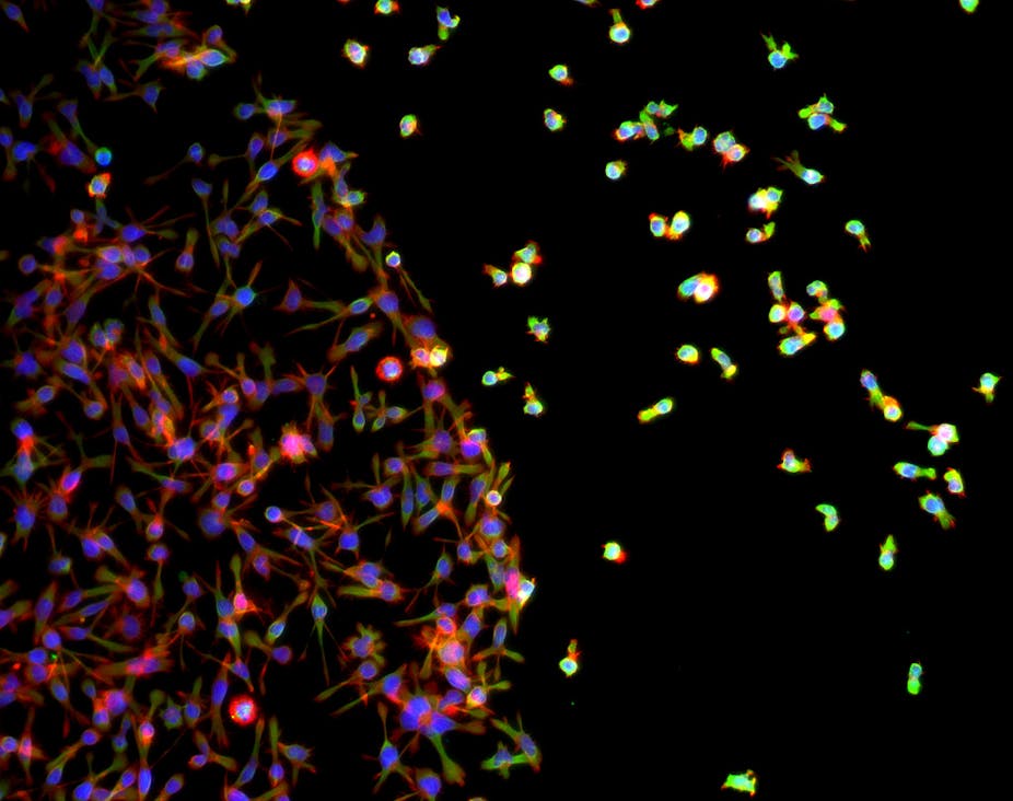
Using the only microscope in Australia capable of super-resolution fluorescence, scientists at the University of NSW were able to identify how exactly the T-cells react – a process they described as a molecular “switch”.
“Previously, it was thought that T-cell signalling was initiated at the cell surface in molecular clusters that formed around the activated receptor,” said lead researcher, Professor Katharina Gaus from UNSW’s Centre for Vascular Research at the Lowy Cancer Research Centre.
“In fact, what happens is that small membrane-enclosed sacks called vesicles inside the cell travel to the receptor, pick up the signal and then leave again,” she said.
“The signalling station is like a docking port or an airport with vesicles like planes landing and taking off. The process allows a few receptors to activate a cell and then trigger the entire immune response,” she said.
Associate Professor Gaus said it was still too early to say exactly how the discovery could help patients with auto-immune diseases or cancer.
“I have had a number of patients calling up this morning asking me this question but we are scientists so this is a long way off from being a diagnostic tool,” she said.
“But if we can identify the machinery that facilitates that recruitment process to the membrane, then we can inhibit that machinery specifically. We can suppress T-cell activation, for example in an auto-immune diseases (such as lupus or Type 2 Diabetes).”
The super microscope allowed researchers to see molecules as small as 10 nanometres and follow what individual molecules were doing, which would be impossible under a regular microscope.
“In conventional microscopy, all the target molecules are lit up at once and individual molecules become lost amongst their neighbours – it’s like trying to follow a conversation in a crowd where everyone is talking at once,” said PhD candidate David Williamson, whose research formed the basis of the paper.
“With our microscope we can make the target molecules light up one at a time.”
“Being able to see the behaviour and function of individual molecules in a live cell is the equivalent of seeing atoms for the first time. It could change the whole concept of molecular and cell biology,” Mr Williamson said.
The research was funded by the National Health and Medical Research Council, the Australian Research Council and Human Frontier Science Program.
The study was published in the journal Nature Immunology.