


The microscope is an iconic symbol of the life sciences – and for good reason. From the discovery of the existence of cells to the structure of DNA, microscopy has been a quintessential tool of the field, unlocking new dimensions of the living world not only for scientists but also for the general public.
For the life sciences, where understanding the function of a living thing often requires interpreting its form, imaging is vital to confirming theories and revealing what is yet unknown.
This selection of stories from The Conversation’s archive presents a few ways in which microscopy has contributed to different forms of scientific knowledge, including techniques that take visualization beyond sight altogether.
1. Seeing as identifying
Over the past few centuries, the microscope has undergone a gradual but significant evolution. Each advance has allowed researchers to see increasingly smaller and more fragile structures and biomolecules at increasingly higher resolution – from cells, to the structures within cells, to the structures within the structures within cells, down to atoms.
But there is still a resolution gap between the smallest and largest structures of the cell. Biophysicist Jeremy Berg drew an analogy to Google Maps: Though scientists could see the city as a whole and individual houses, they couldn’t make out the neighborhoods.
“Seeing these neighborhood-level details is essential to being able to understand how individual components work together in the environment of a cell,” he writes.
Scientists are working to bridge that resolution gap. Improvements to the 2014 Nobel Prize-winning superresolution microscopy, for example, have enhanced the study of lengthy processes like cell division by capturing images across a range of size and time scales simultaneously, bringing clarity to details traditional microscopes tend to blur.
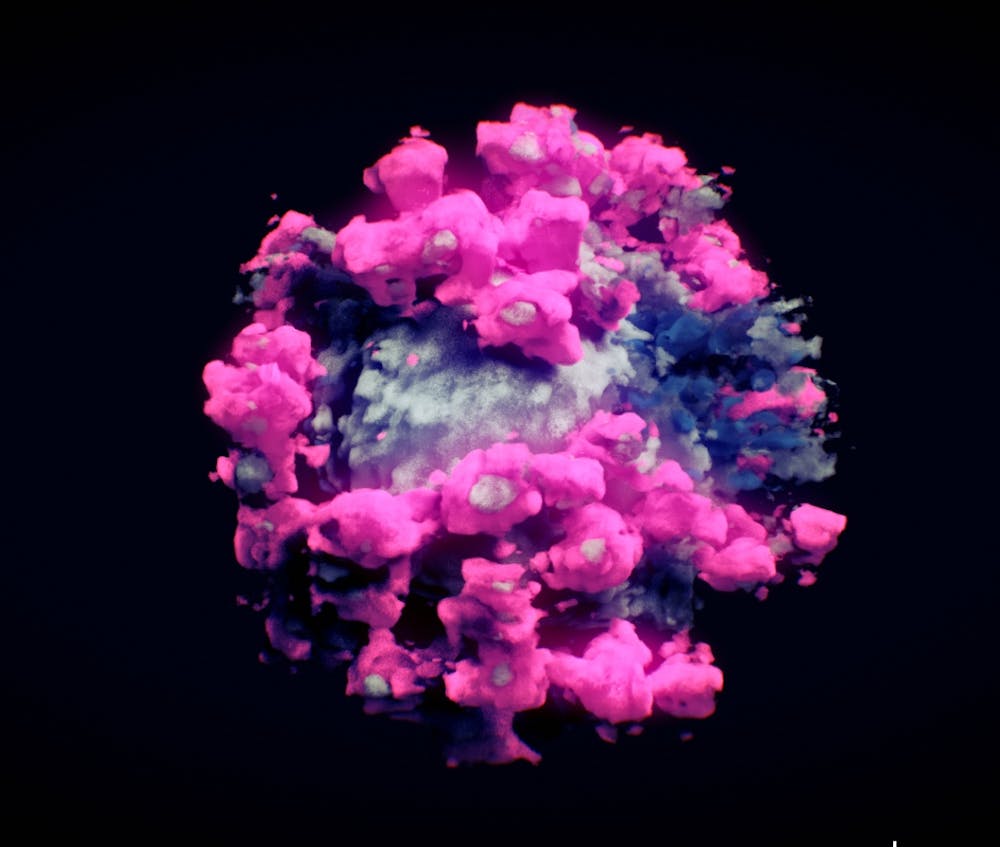
Another technique, cryo-electron microscopy, or cryo-EM, won a Nobel Prize in 2017 for bringing even more complex, dynamic molecules into view by flash-freezing them. This creates a protective glasslike shell around samples as they’re bombarded by a beam of electrons to create their photo op. Cryo-ET, a specialized type of cryo-EM, can construct 3D images of molecular structures within their natural environments.
These techniques not only generate images at or near atomic resolution but also preserve the natural shape of difficult-to-capture biomolecules of interest. Researchers were able to use cryo-EM, for instance, to capture the elusive structure of the protein on the surface of the shape-shifting hepatitis C virus, providing key information for a future vaccine.
Further enhancements to science’s visual acuity will reveal more of the fine details of the building blocks of life.
“I anticipate seeing new theories on how we understand cells, moving from disorganized bags of molecules to intricately organized and dynamic systems,” writes Berg.
2. Seeing as scoping
Microscopy images are often framed as snapshots – circumscribed parts of a whole that have been magnified to reveal their hidden features. But nothing in an organism works in isolation. After discerning individual components, scientists are tasked with charting how they interact with each other in the macrosystem of the body. Figuring this out requires not only identifying every component that makes up a particular cell, tissue and organ but also placing them in relation to each other – in other words, making a map.
Researchers have been charting the brain by stitching together multiple snapshots like a photo mosaic. They use different techniques to label a specific cell type and then image the whole brain at high resolution. Layer by layer, each run-through creates an increasingly detailed and more complete model. Neuroscientist Yongsoo Kim likens the process to a satellite image of the brain. Combining millions of these photos allows researchers to zoom into the weeds and zoom out to a bird’s-eye view.
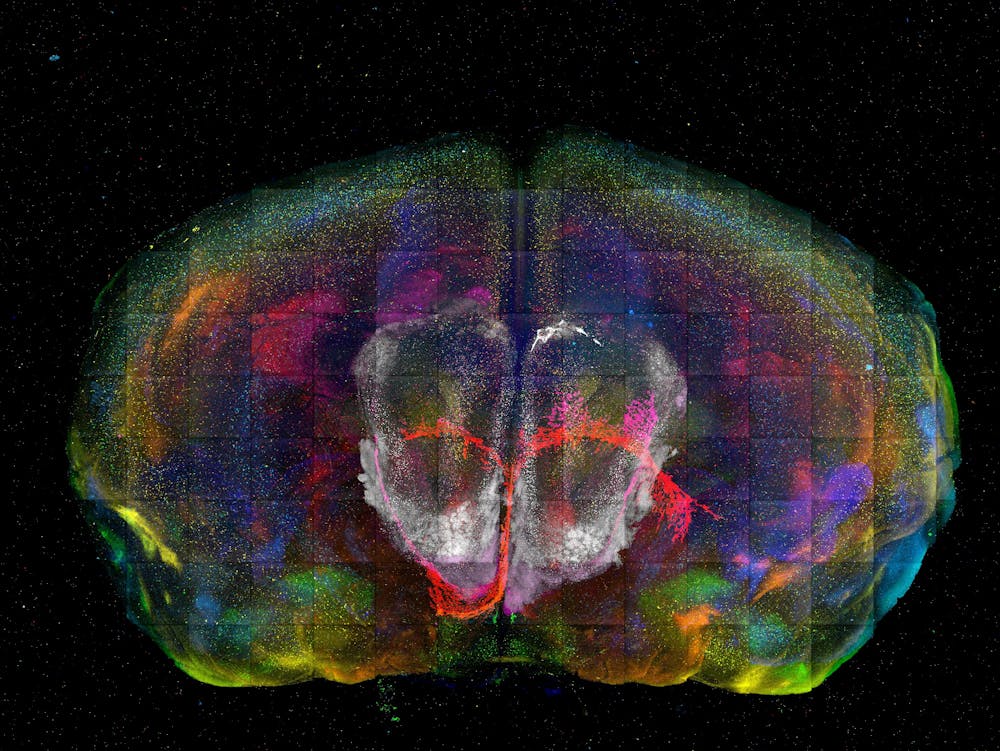
But building a map of a city, however detailed, is not the same as understanding its rhythm and atmosphere. Likewise, knowing where every cell is located relative to each other doesn’t necessarily tell researchers how they function or interact. Just as important as charting out the landscape of an organ is coming up with a working theory of how it all fits together and performs as a whole. Right now, Kim notes, analysis lags behind technical advances in data collection.
“Incredibly rich, high-resolution brain mapping presents a great opportunity for neuroscientists to deeply ponder what this new data says about how the brain works,” Kim writes. “Though there are still many unknowns about the brain, these new tools and techniques could help bring them to light.”
3. Seeing as recognizing
Every improvement in technology brings a parallel improvement in the data it collects, both in quality and in quantity. But that data is only useful insofar as researchers are able to analyze it – high granularity isn’t helpful if those details aren’t appreciable, and high output isn’t beneficial if it’s too overwhelming to organize.
Automated microscopes, for example, have made it possible to take time-lapse images of cells, resulting in massive amounts of data that require manual sifting. Neuroscientist Jeremy Linsley and his team encountered this dilemma in their own work on neurodegenerative disease. They’ve been relying on an army of interns to scour hundreds of thousands of images of neurons and tally each death – a slow and expensive process.
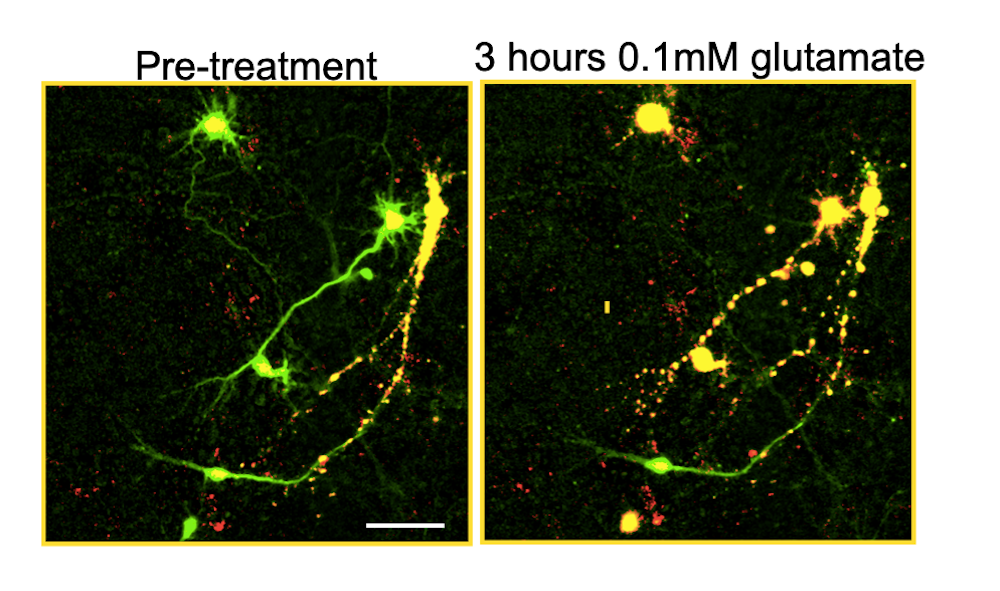
So they turned to artificial intelligence. Researchers can train an AI model to recognize specific patterns by feeding it many sample images, pointing out structures of interest and extrapolating the algorithm to new contexts. Linsley and his team developed a model to distinguish between living and dead neurons with greater speed and accuracy than people trained to do the same task.
They also opened the black box of the model to figure out how it was finding dead cells, revealing new signals of neuron death that researchers previously weren’t aware of because they weren’t obvious to the human eye.
“By taking out human guesswork, (AI models) increase the reproducibility and speed of research and can help researchers discover new phenomena in images that they would otherwise not have been able to easily recognize,” writes Linsley.
4. Seeing as appreciating
Even before they had the instruments to zoom in on samples, researchers had a tool in their arsenal to study the living world that they still use today: art.

Centuries ago, scientists and artists examined plants, animals and anatomy through illustration. Sketches of unfamiliar species in their natural environments aided in their classification, and drawings of the human body advanced study of its structure and function. With the help of the printing press, these artistic renderings – which later included the view under the lenses of early microscopes – popularized scientific knowledge about the natural world.
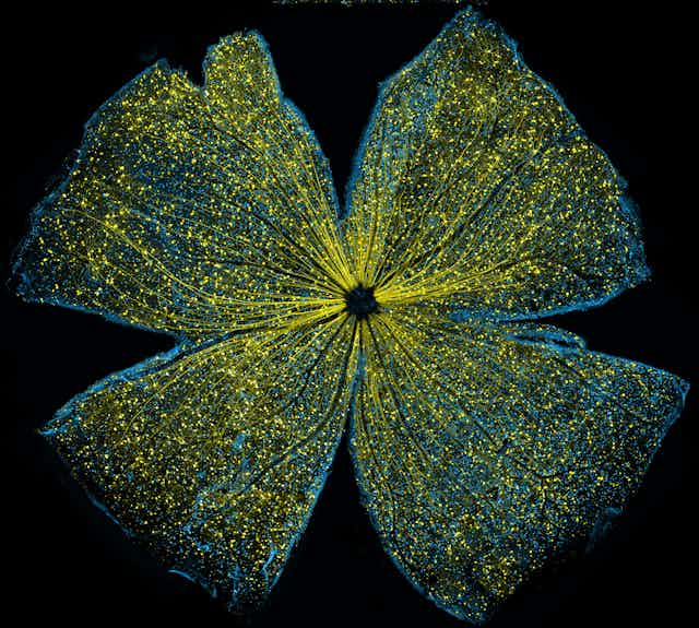
Though hand drawings have since given way to advanced imaging techniques and computer models, the legacy of communicating science through art continues. Scientific publications and BioArt competitions highlight laboratory images and videos to share the awe and wonder of studying the natural world with the general public. Using visualizations in classrooms and art museums can also promote science literacy by giving students a chance to look through the eye of the microscope as a scientist would.
Biologist and BioArt Awards judge Chris Curran believes that making visible the processes and concepts of science can grant a greater depth of understanding of the natural world necessary to being an informed citizen.
“That those images and videos are often beautiful is an added benefit,” she writes.
And the abstract qualities of science can be made tangible in ways that don’t involve sight. Proteins, for instance, can be translated into music by mapping their physical properties into sound: amino acids turn into notes, while structural loops become tempos and motifs. Computational biologists Peng Zhang and Yuzong Chen enhanced the musicality of these mapping techniques by basing them on different music styles, such as that of Chopin. Consequently, a protein that prevents cancer formation, p53, sounds toccata-like, and the protein that binds to the hormone and neurotransmitter oxytocin flutters with recurring motifs.
Framing scientific images as art often requires no more than a change in perspective. And uncovering the poetry of science, many researchers would agree, can help reveal the artistry of life.