Nature, red in tooth and claw.
When Tennyson published his poem In Memoriam, little did he know that this phrase from it would become so intimately associated with the process of Darwinian natural selection. Five little words which evoke the harsh evolutionary realities of competition for food, resources and life itself between predator and prey, the hunter and the hunted.
Now my colleagues and I, led by Lida Xing from the China University of Geosciences (Beijing), have published evidence of one lucky animal that got away – in this case, a herbivorous dinosaur from China. Our work highlights how the use of X-ray tomography – a rapidly developing technique in digital imaging – is revolutionising the study of the fossil record.
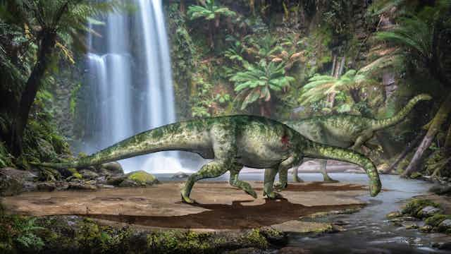
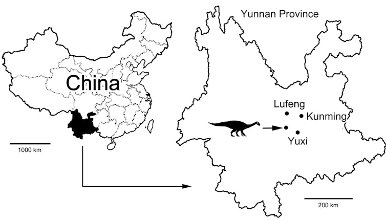
Our dinosaur is Lufengosaurus huenei, a Lower Jurassic sauropod, who would have lived 200-170m years ago in what is now Yunnan Province, China. Lufengosaurus was a herbivore, around six metres in length and weighing a little under two tonnes.
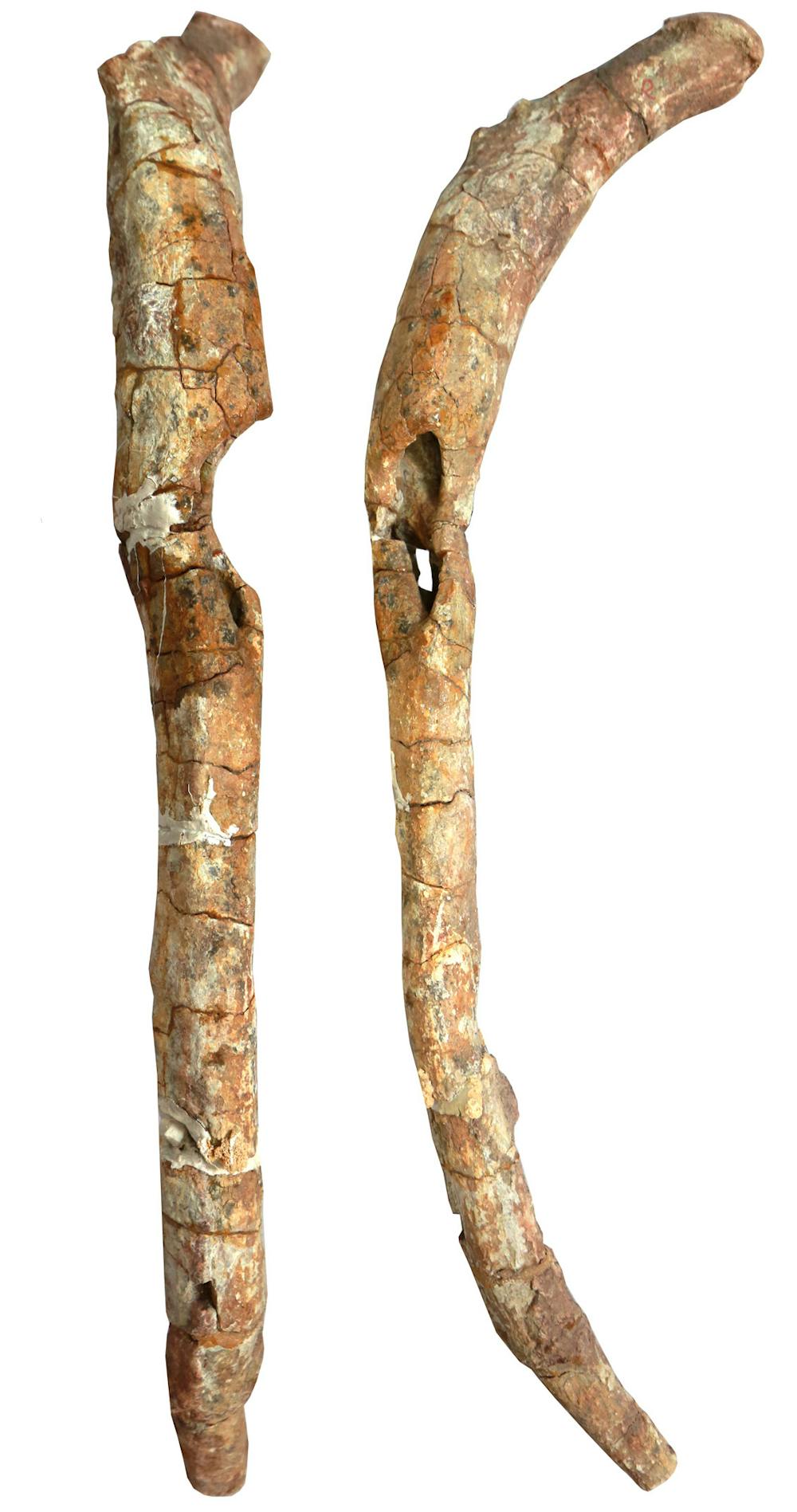
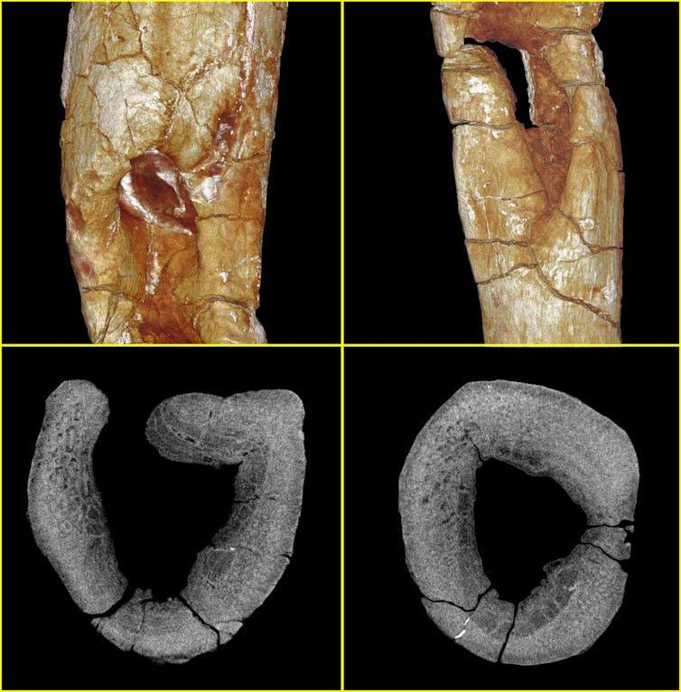
When the dinosaur was excavated in 1997, there was a pathological abnormality on one of the right ribs of the animal. Viewed from the side, there is a concave section of missing bone which cuts almost halfway through the rib.
The traditional approach in studying bone pathology is what is termed “morphoscopic evaluation”. This usually involves low powered magnification of the bone, but this would only image the external surface of the fossil. In the case of our rib, the lesion penetrated deep into the bone, so seeing the internal structure was needed for a diagnosis.
Now, 20 years after its initial discovery, we have used X-ray micro-computed tomography, or micro-CT for short, to image the deep structures of our dinosaur.
Seeing inside fossils
Tomography (from the Greek tomos to slice, and graphos to write) is a non-invasive technique that has significant diagnostic advantages over conventional methods, allowing high-resolution slices and 3D images to be built up of internal structures without damaging the fossil.
Following micro-CT scanning, we reconstructed the cellular structure of the rib. In cross-section, there was clear evidence of both destructive changes and new bone formation which could not be observed from the outside. The pattern of these bone-destroying and bone-forming processes tells us that the disease process was both chronic (long-term) and active at the time of the animal’s death.
We diagnosed a process called osteomyelitis, which in this case had produced an abscess inside the bone. Osteomyelitis is a severe infection originating in the bone marrow, usually resulting from the introduction of pyogenic (pus-producing) bacteria into the bone. Pathogens enter the bone via the bloodstream, or through open wounds or fractures.
This is only the second case of osteomyelitis to be found in a sauropod dinosaur in the fossil record. The only other case comes from a giant titanosaur from Argentina who had a bacterial infection of the spine.
Tooth and claw
In this Lufengosaurus we also have the earliest recorded case of a bony abscess caused by osteomyelitis in the fossil record.
Given the shape of the lesion, and its position on the ribcage, we think that the infection may have been caused by a puncture wound from a bite. The teardrop shape suggests that the damage was produced by a tooth or claw, and is in keeping with evidence for predator bite trauma found elsewhere in the dinosaur fossil record.
The bacterial infection would have had a big impact on the life of the Yunnan dinosaur. Osteomyelitis is known to produce fever, fatigue, nausea and discomfort, and may send tracts of bacteria into the brain, accelerating death. We know that the dinosaur survived for some time with this infection, but this may have made it vulnerable to other diseases or unable to fend for itself in the long term.
What is exciting is that this case gives us evidence of interaction between a large plant-eating dinosaur (a sauropod) and one of the aggressive predators living at that time. We don’t just have evidence of disease but of behaviour between animals – between predator and prey at this deep period in prehistory.
We do not know which species of predator caused the bite, but the wound from the failed attack is a smoking gun. It is possible that Sinosaurus, a well-known predator found in Jurassic Yunnan, would have been able to attack Lufengosaurus.
Virtual palaeontology
This discovery was only made possible by the application of X-ray tomography (micro-CT). The first commercially available micro-CT scanner appeared in 1994, but it is only in the last decade that it has begun to be used in palaeontology, partly because of the cost of the equipment. Tomography is increasingly allowing us to understand processes such as trauma and infection in the fossil record at the cellular level.
This technology has opened up the fossil record, allowing palaeontologists to image and analyse the deep structure of fossils. This has enabled spectacular discoveries such as the earliest hominin cancer and the earliest tumour, the flight pattern of Archaeoptryx, or to rebuild an early bird trapped in amber. It has also allowed us to correct historical cases of pathological misdiagnosis in fossils.
The resulting scans can be shared across the world, visualised and studied without the need to access the fossils directly. They can also be 3D printed, both in their actual size or at any other scale that we require.
Who knows what spectacular discoveries await us using this technology, but it is clear that the future of palaeontological research is virtual.