
This article is published in collaboration with researchers of the Institut de Systématique, Évolution, Biodiversité (ISYEB) of the Muséum National d'Histoire Naturelle, Sorbonne Universités, Paris.
Do you know about “comparative spermatology”? It’s the science of describing spermatozoa. A first international congress was devoted to it in 1970. In 1976, more than 1,000 animal species had their spermatozoa described by electron microscopy; and today it’s probably closer to 10,000.
To understand evolution, scientists start by observing living things, and by defining their characteristics. For example, the number of legs, the presence of feathers. We then need to unravel these characteristics and to understand which ones make it possible to trace the evolutionary lines.
In the history of biology, increasingly powerful instruments have been used to describe and classify these characteristics: from Antiquity people used the naked eye, then magnifying glasses, microscopes, and finally electron microscopes. For about the last thirty years, the tools of molecular biology have given us access to the four bases of DNA (A, T, G, and C), which are also characteristics.
For some animals, it was very difficult to understand evolution from observations. This was particularly the case for parasitic worms belonging to the Phylum Platyhelminthes (or flatworms). These animals are soft and have no fossils. Their flat shape and the apparent simplicity of their anatomy did not give many clues to understand their evolution. In other words, they lacked distinguishing characteristics.
Spermatozoa diversity
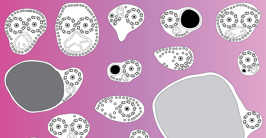
In the 1970s, the electron microscopy revolution made it possible to rediscover the incredibly variable world of the shape of cells and their organelles. Cell biology then, once again, became descriptive. And among these cells, one appeared extremely variable: the spermatozoon. The spermatozoon plays the very specialised role of transporting the genetic information of the male and bringing it to the female cell, the egg. The egg is a spherical cell without much originality in most animals. But the spermatozoon! What an incredible variety of shapes and sizes!
Let’s go back to our parasitic worms, members of the Platyhelminthes. Although they have a soft and uninteresting body, there is incredible diversity in their sperm! It must be said that a parasite, comfortably installed in its host, which provides food and shelter, has only one thing to do: to reproduce. Lay millions of eggs, make millions of oocytes, and more millions of spermatozoa, to create a new generation of parasites. To understand the evolution of these worms, one way is for researchers to study their spermatozoa.
What differentiates a human sperm from that of a flatworm? Axonemes, among other features. These structures are the motors of cilia and flagella in eukaryotic cells. We humans have cilia in many cells, such as in the bronchi, and flagella in our spermatozoa. In 99% of eukaryotic cells, these axonemes have the same organization, with 9 doublets of microtubules in a circle and two central microtubules; this almost universal structure is called 9 + 2. The component of these microtubules is a protein, tubulin.

But in the Platyhelminthes, the axonemes of the spermatozoa do not have this universal 9 + 2 structure. In their center, they do not have microtubules but a spiral core; these axonemes are therefore called 9 + "1". Note the quotation marks, because the “1” in the middle is not half of 2 of the 9 + 2 axonemes: it is something completely different. A different structure in the center of the axoneme: now that’s a characteristic!
And this is precisely what becomes useful for understanding evolution: this bizarre 9 + "1" axoneme is shared by all the parasitic Platyhelminthes and also some groups of non-parasitic Platyhelminthes, which belong to the so-called Turbellaria. Thanks to this characteristic, as early as 1985, Ehlers was able to retrace the evolution of the Platyhelminthes and bring some of them together in a group he called the Trepaxonemata (from trepa which means “in spiral”: those that have a spiral in their axoneme). Other more detailed work has found other characteristics in Platyhelminth spermatozoa: for example, the number of axonemes. Most spermatozoa of Platyhelminthes have two axonemes (a rarely encountered situation in nature), but some have only one.

In the 1990s, comparative spermatology of Platyhelminthes provided researchers with quantities of characteristics, which they lacked before. Thus, Cestodes, or tapeworms, can be defined on the basis of a simple characteristic of their spermatozoa. In the group of Trematodes, which include the flukes and schistosomes, spermatozoa are now known in over a hundred species and also provide useful characteristics for understanding the relationships between certain families.
For the group of Monogenes, which are parasites of fish, the spermatozoa allowed us to qualify, by their characteristics, the two large groups that compose the Monogenes. These groups bear the rather long names of Monopisthocotylea and Polyopisthocotylea, which become almost clear when we understand that they mean “a single sucker at the back” (mono – opistho – cotylea) or “several suckers at the back” (poly – opistho – cotylea). Let’s call them “Polyops” and “Monops” for simplicity.
How to distinguish Polyops from Monops

Well, we can recognize a Polyop from a Monop – thanks to its sperm. Those of the Polyops have two axonemes and microtubules on the sides, while the different families of Monops have different kinds of spermatozoa, which are never like those of Polyops. There remains a problem: nothing in sperm allows us to recognize a Monogene – in other words, the Polyops and Monops form a group, each on their own (each group descends from a common ancestor), but nothing brings them together (there is no ancestor common to all Monogenes). Or, in scientific terms, the characteristics of sperm prove that Polyops and Monops are monophyletic, but the Monogenes, in principle formed by the addition Monops + Polyops, are not.
When we obtained the first results from molecular biology, the evolutionary trees calculated from these new characteristics (the bases A, T, G, and C) quickly gave the same results, with Monops and Polyops monophyletic, but never gave us evidence of the monophyly of the Monogenes.
Now we have published new results on Polyop spermatozoa. Why not do that earlier? Because it’s not so easy. Monogenes are parasites of cartilaginous and bony fish. The most primitive Monogenes are parasites of cartilaginous fish (Chondrichthyans), the chimaeras and sharks. And it is not easy to fish out a chimaera, which lives in the bottom of the oceans, in good condition, with its parasites very fresh.

New results
We obtained results on spermatozoa of three rare Polyops: two of the family Hexabothriidae (the name means that they have six – hexa – suckers – bothrium). And above all, one of the family Chimaericolidae (a name that is easy to remember as it is a chimaera parasite), whose species name is Chimaericola leptogaster; we collected this parasite from a chimaera fished off Norway. Nobody had yet observed the spermatozoon structure of these two families. Surprisingly, while these two families are undoubtedly Polyops (they have several suckers in the back, this is clearly visible), their spermatozoa do not have the structure of Polyop sperm. This is a 20-year certainty that is disappearing, and this shows that it is not only molecular biology that can still bring new results. Finally, the characteristics of the spermatozoa that united, it seemed, Polyops, are restricted to a terminal branch of the Polyops (the Mazocraeidea), which are parasites of bony fish (Osteichthyes).

By the way, what do the recent results of molecular biology say? The latest trend is to sequence the entire genome of the mitochondria and compare, not just the ATGC base sequences, but rather the order of the genes and various details in this mitochondrial genome, the mitogenome, which is circular. The characteristics are no longer the bases themselves but the arrangements of the groups of bases. What do the mitogenomes of the Monogenes say? That the Polyops are similar between themselves, that the Monops are similar between themselves … but that the Polyops do not look like the Monops.
And that brings us back to the results of comparative spermatology 20 years ago. But what about the mitogenomes of Chimaericolidae and Hexabothriidae? No data available! The Polyops for which we know the mitogenome are all Mazocraeidea, the parasites of bony fish mentioned above. Our knowledge of the mitogenomes of the Monogenes is at the point where the knowledge of the spermatozoa was 20 years ago. See you in a few years…