
Melanocytes are a small subset of epidermal cells that play an outsize role in protecting your skin from the damaging effects of sun exposure. They do this by synthesizing melanins, which are pigments sent to other skin cells to shield them from harmful ultraviolet light. A lack of functioning melanocytes causes a wide range of skin conditions, including skin cancer and vitiligo, an autoimmune condition in which the body attacks melanocytes and causes patches of depigmented skin.
For nearly 20 years, I have been studying melanocytes and the role they play in disease. Difficulties growing human melanocytes in cell cultures have led researchers like me to use alternative models to study them.
My lab and others have pioneered the use of zebrafish to study melanocytes. Using this small freshwater fish as a model organism, my team and I recently discovered a new way in which melanocytes regenerate. This process enables flexibility for these cells to recover from injuries and may be applicable to other types of tissues.
What zebrafish and people have in common
New students and nonscientists often ask me, “Why zebrafish?” There are several reasons why zebrafish are good models to study melanocytes.
Melanocytes in zebrafish are similar in many ways to those in people. These cells develop in embryos in the same way those in humans do, use the same genetic programs and make the same melanins. Melanocyte dysfunction in zebrafish also leads to the same diseases and cancers found in people.
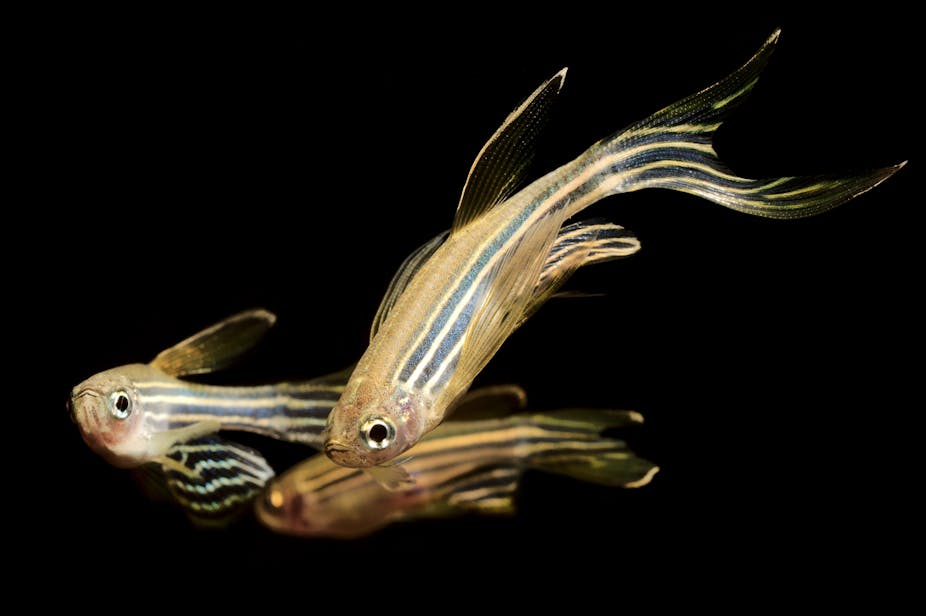
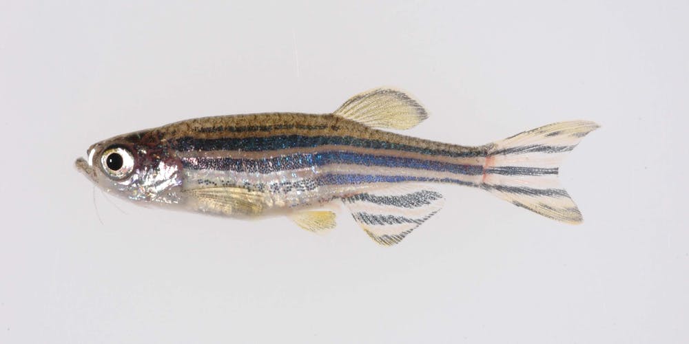
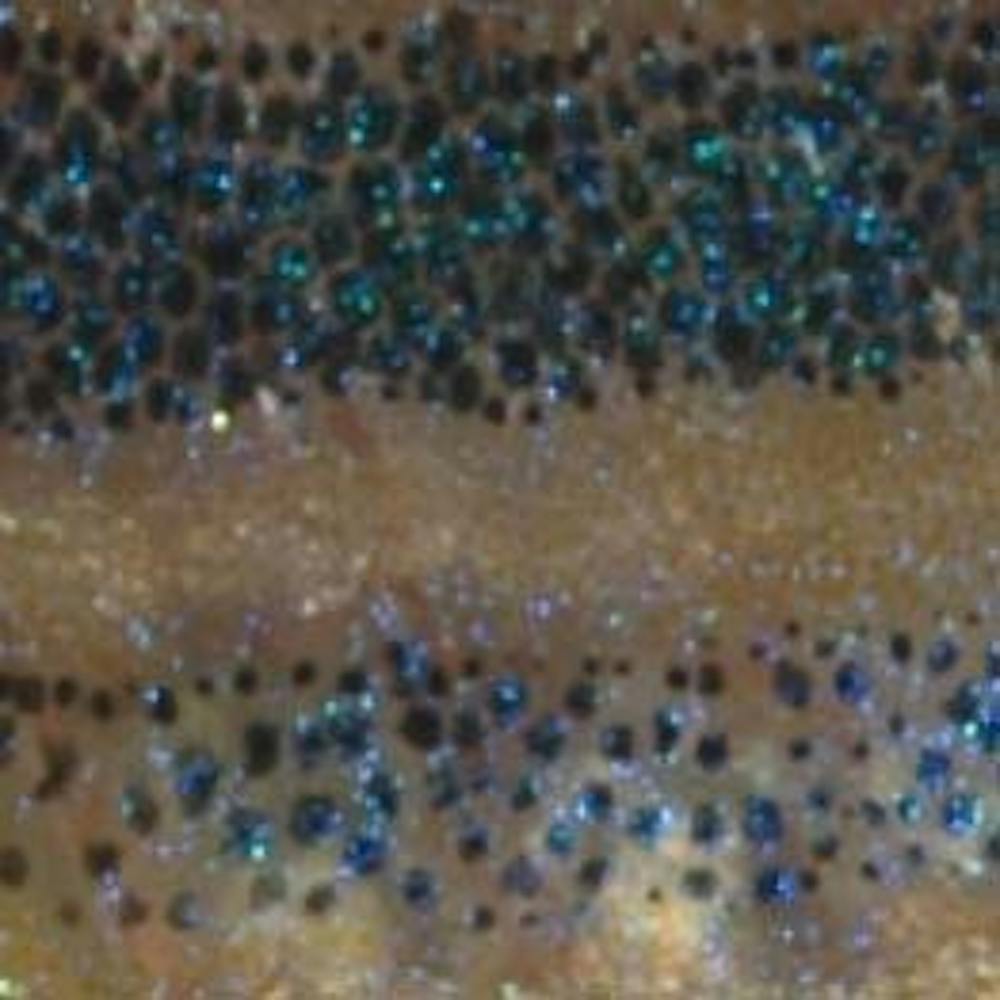
Unlike melanocytes in mouse or human skin, zebrafish melanocytes are externally visible in their dark stripes and spotted scales. Researchers can place the whole fish directly under a microscope and see the cells without the need for a biopsy.
Importantly, researchers can manipulate and perform experiments on zebrafish melanocytes in ways that are unethical or not feasible to do with people. Unlike studies that use isolated melanocytes in a petri dish, these experiments can take place in the context of a whole animal, where we can monitor the surrounding skin and other biological factors for their influence on how melanocytes behave and function.
Diversity of melanocyte stem cells
In work spearheaded by Tyler Frantz, a graduate student in my lab, our team has focused our attention on the process by which new melanocytes regenerate after injury.
Melanocyte regeneration is important for recovering from skin disorders such as vitiligo. It’s also relevant to age-related conditions like hair graying, in which melanocyte stem cells either die or become dormant and no longer produce the mature melanocytes that give hair its color.
To study melanocyte regeneration, we removed these cells from zebrafish and followed their process of regrowth. Since melanocyte stem cells in zebrafish are externally visible, we tracked these cells in real time to see how they divided and matured. Additionally, we measured which genes were expressed in individual melanocyte stem cells and their descendants during regeneration.
We found that dying melanocytes trigger this regenerative process by sending the signal for melanocyte stem cells – cells that can give rise to new melanocytes – to activate. Surprisingly, we identified two types of stem cells that each took a different route to make new melanocytes. One type of stem cell directly converted into melanin-producing melanocytes. The other type of stem cell divided to create two types of daughter cells. One type was new melanocytes, and the other was new stem cells ready to respond to future injury.
Researchers have known that a single stem cell is capable of making the multiple types of cells needed to regenerate tissue. Our zebrafish studies indicate that multiple different stem cells in skin, and potentially other tissues, can together reconstruct one particular cell type after injury. The involvement of multiple stem cells likely enables regeneration to nimbly adjust to different types of injuries.
From fish to people
Our findings from zebrafish are likely relevant to human skin. When we examined cells taken from the fluid within a blister in human skin, we found cells that look remarkably similar to zebrafish melanocyte stem cells. We are planning to see whether these human cells are activated in skin regeneration to make new melanocytes, which would confirm their identity as melanocyte stem cells.
Ultimately, we envision using these findings to develop treatments that reinvigorate melanocyte stem cells, which could help reverse skin color loss in vitiligo and other diseases. Such treatments may also help counteract age-related pigment loss in hair and skin.
The unique features of zebrafish have allowed us to uncover a new mode of cellular regeneration. Because of cross-species similarities, we expect that these and many other findings from research using zebrafish may be applied to human biology.