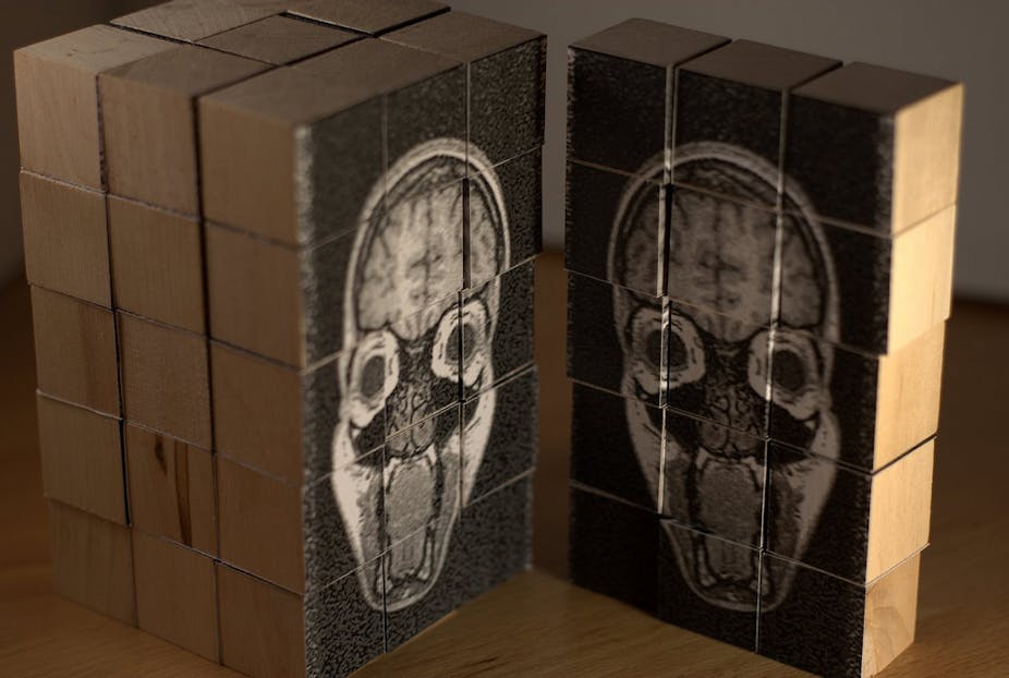
A serious illness or brain injury can put a person into a coma: a sleep-like or a vegetative state where they are awake but unaware. These disorders of consciousness are sometimes not accurately diagnosed because doctors rely on behavioural cues to make decisions, but these cues only provide a limited perspective of a person’s capacities for consciousness.
Included in these disorders is the “minimally conscious state”, where a patient can exhibit signs of voluntary responsiveness, simple movements like lifting a finger. If patients do show signs of responsiveness, despite limited mobility, we can use other ways to try and communicate with them.
Causing a stir
In 2006, it was demonstrated by Adrian Owen and colleagues that brain imaging using functional magnetic resonance imaging (fMRI), which measures activity by detecting changes in blood flow in the brain, could produce similar brain activation patterns in someone in a vegetative state to healthy controls when they were asked to imagine navigating their way around their home or playing tennis.
This result was for a single patient, but nevertheless caused a stir in both the medical and research communities. If a patient is diagnosed as vegetative but can perform mental imagery tasks as evidenced by brain imaging, then was the original diagnosis wrong?
The first question is whether the brain activations seen during mental imagery tasks correspond to voluntary conscious or automated unconscious information processing. This is difficult to answer because conscious experience is subjective and, therefore, only the patient can know if they were performing the task voluntarily.
Since this first study, many groups around the world (including ours) have been doing similar studies to both validate and further understand possible ways of extending communication with patients in various states of consciousness. The ultimate aim is to apply such methods in clinical practice and treatment. A general finding has been that not all patients are able to produce brain activation patterns during mental imagery tasks that are similar to controls.
Traumatic and acquired injury
In a larger study by the groups of Adrian Owen and Stephen Laureys, it was found that five in 54 vegetative state or minimally conscious patients could perform at least one mental imagery task using imaging methods.
This group can be divided into patients with traumatic brain injury (resulting from an accident or an assault for example) and acquired brain injury (resulting from a condition such as stroke or lack of oxygen to the brain).
Of the traumatic brain injury patients, five of 33 could perform at least one mental imagery task, while none of the acquired brain injury patients could perform the task. Similar findings were obtained in a study performed by our group. The limited success with acquired brain injury patients may be linked to more diffuse damage in brain areas important for producing consciousness compared with those who’ve suffered a traumatic brain injury.
Voluntary or automation
“Positive” brain activation is indicative of functioning brain tissue, but not necessarily voluntary, conscious information processing. So although we may see positive brain activations in some patients and not others using imaging methods, we’re not yet at the stage where this information can be used to modify a patient’s treatment. Similarly, if a patient does not show brain activation on a medical imaging scan, it does not necessarily mean the patient’s brain isn’t functioning or that the patient isn’t aware, although reduced brain activations are a sign of limited brain function. It also may be that the patient can’t perform a particular task but may be able to perform others.
Key problems include the variability in patient state and brain damage as well as the level of “noise” inherent in the imaging techniques - for example scanner-induced artefacts or (involuntary) patient movement sensed in the recorded signals. We need to improve this signal-to-noise ratio to determine what signal patterns mean in terms of conscious state.
Another important issue in this field of research is the cost of the imaging techniques. An fMRI scanner can cost millions of dollars and is completely immobile. This means patients need to be brought to the scanner in order to perform studies so they can’t be done on a daily basis, which is important for regular interaction and stimulation of the patient and is known to play an important role in the outcomes of patients with disorders of consciousness.
Electromyography (EMG, measurement of electrical muscle activity), which is being used to detect potential voluntary movements in people with consciousness disorders, and electroencephalography (EEG, measurement of electrical brain activity), also being used with a varying degree of success to detect awareness, can overcome this accessibility problem because the recording systems involved are much more affordable and portable.
But although these techniques have a superior temporal resolution to fMRI, they don’t provide the perfect solution because their spatial resolution is inferior.
We know that observing behaviour alone will not give us a full picture of what is happening in the brain in people in vegetative states and other states of consciousness. But imaging might provide a way of communicating beyond the body. And fMRI and other complementary imaging techniques are all likely to play a role in future forms of clinical treatment.
The brain is incredibly complex and there is much that we still don’t know. But working out what may or may not be a voluntary action will help us determine next steps for reaching out and communicating with people with some of these consciousness disorders.

