
“Medical illustrators draw what can’t be seen, watch what’s never been done, and tell thousands about it without saying a word.”
For decades, this slogan appeared on the website and printed materials of the Association of Medical Illustrators. Although the association no longer uses this tag line, it’s still an accurate description of the profession.
As a practicing medical illustrator for over 30 years, I draw what can’t be seen and watch what’s never been done on a daily basis. And I teach my students to do the same.
But what exactly does all of that mean, and how does it improve medicine?
Tell thousands about it without saying a word
You may have heard the adage, “A picture is worth a thousand words.” In that same vein, medical illustrators use pictures to teach complex scientific concepts. As the famed medical illustrator Frank H. Netter once said, “(Pictures) eliminate the need for the lecturer or the author to translate what he has in his mind into words and for the listener or the student to translate those words back into a mental image.”
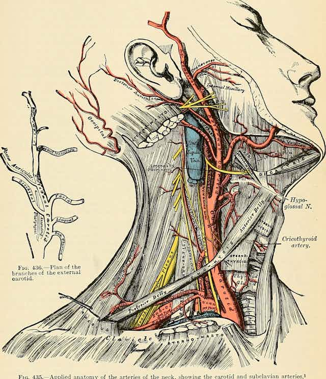
The use of illustrations to communicate medical information has a long history, dating back at least to ancient Egypt and flourishing in the Renaissance. The work of 16th century anatomists Giacomo Berengario da Carpi and Andreas Vesalius set a precedent for the use of detailed illustrations to teach anatomy, a practice that continues to this day.
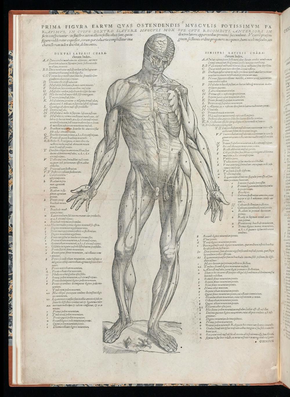
The proliferation of illustrated anatomy atlases in the Renaissance coincided with the widespread acceptance of cadaver dissection. The earliest known human dissections were performed in the third century BCE. The practice was prohibited throughout the Middle Ages but became common again in the 13th and 14th centuries.
By the 1500s, dissections, usually of executed criminals, had become public spectacles. The demand for bodies eventually outstripped the supply of executed convicts, leading to the unscrupulous practices of grave robbing and even murder.
In addition to depicting the location and features of an object such as an organ, illustrations proved essential in describing events happening over time, such as the progression of a disease or the steps in a surgical procedure. Generations of surgeons learned new procedures from meticulously illustrated surgical atlases. An early example of physiology illustration, William Harvey’s classic 17th century work on the circulation of blood, “Exercitatio Anatomica de Motu Cordis et Sanguinis in Animalibus,” depicts the direction of blood flow through the veins of the forearm.

_Venenbild.jpg)
Nowadays, surgeons can practice a procedure hundreds of times in virtual reality before trying it on a real patient. Modern physiology and pathology texts include countless illustrations of the body, not just at the anatomical level but also the cellular and molecular. So valuable are these depictions of complex pathways and interactions that many science journals now require papers to include a graphical abstract, a single illustration that summarizes the content of each paper.
Draw what can’t be seen
Medical illustrators employ special tools and training to visualize things that are normally hidden from the naked eye.
All professionally trained medical illustrators study human gross anatomy, including dissecting a human cadaver, in order to visualize the internal structures of the body. When a cadaver isn’t readily available to serve as reference for an illustration, illustrators use medical imaging, such as CT and MRI scans, and reconstruct the body in three dimensions.
At the cellular level, medical illustrators must understand how to use microscopy techniques in order to find references for accurate depictions of cellular structures.
Objects at the smallest scale – atoms and many molecules – are smaller than the wavelength of visible light. This means they are below the theoretical limit of what can be seen, even with the most powerful light microscope. So researchers experimentally determine the structures of molecules using techniques like X-ray crystallography and nuclear magnetic resonance spectroscopy instead. These techniques use X-rays or radio waves, respectively, to determine how atoms are arranged.
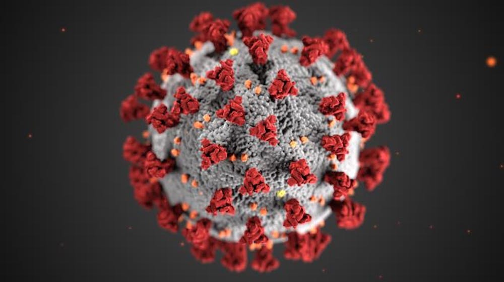
Medical illustrators learn to locate and retrieve data on the structure of molecules from sites like the RCSB Protein Databank. They also use a host of visualization applications and software plug-ins to render these structures in 3D.
Medical illustrators Alissa Eckert and Dan Higgins at the U.S. Centers for Disease Control and Prevention used these techniques to create the famous red-spiked coronavirus image that went viral during the pandemic.
Watch what’s never been done
Obviously, you can’t really watch something that has never been done. But medical illustrators can help conceptualize new processes and techniques before they become a reality.
For example, they might illustrate how an experimental drug may theoretically work before it enters testing. Similarly, illustrations can be critically important in pre-surgical planning, especially in complex cases.
My favorite example of the role of medical illustration in surgery is the separation of conjoined twins Abbigail and Isabelle Carlsen at the Mayo Clinic in 2006. Working from nearly 6,000 radiographic images, the clinic’s medical illustrators produced five detailed illustrations of the twins’ anatomy. They even generated 3D-printed models of important structures, notably their shared liver.
The illustrations were critical in training a team of 70 surgeons, nurses and technicians involved in the case. They also served as a road map for the ultimately successful surgery, hung up on the walls of the operating theater during the procedure.

Road to becoming a medical illustrator
In order to draw what can’t be seen and watch what’s never been done, medical illustrators require specialized training. Most medical illustrators in North America are trained at master’s programs accredited by the Association of Medical Illustrators in conjunction with the Commission on Accreditation of Allied Health Education Programs.
Since the profession requires a strong understanding of the biomedical sciences, students accepted into these programs must have a strong science background along with a portfolio demonstrating outstanding drawing skills. Students often have a double major in biology and art or a major in one area and minor in the other.
Once in the program, their science training continues with human gross anatomy and some combination of courses in neuroanatomy, embryology, histology, cell biology, pathology and immunology. Specialized courses in surgical observation and cellular and molecular visualization also include significant science content.
Students receive extensive training in computer graphics, including 2D digital illustration and animation, 3D computer modeling and animation, interactive media, virtual and augmented reality and educational game and mobile app design. Courses also emphasize the principles of design, including the use of color, layout and motion to create effective visuals.
Medical illustrators learn to consider the educational level of their audience, since their work may be used to educate patients – even kids – in addition to medical professionals. Illustrations made for a child recently diagnosed with leukemia would be very different from those aimed at the oncologist treating the disease.
After entering the workforce, many medical illustrators pursue optional board certification to become a certified medical illustrator, which recognizes professional competency and encourages continued learning. Continued certification requires 35 hours of continuing education every five years in the biomedical sciences, artistic techniques and business practices.
All of this education and training is essential to ensure that medical illustrators communicate complex scientific information with accuracy and clarity. I like to think of medical illustrators as teachers – they instruct with pictures.