
Our series, Animals in Research, profiles the top organisms used for science experimentation. Here, we look at Caenorhabditis elegans – a roundworm.
When you think of a worm, what do you see? For some it’s the squeamish thought of treading on one in bare feet after rain; for others it’s the periodical doses of dewormer for the family pet. Perhaps it’s even the slimy tubes that happily munch through the compost.
Well, now you can add “medical research star” to your list. One species of worm - Caenorhabditis elegans - has contributed more to medical science in the past few decades than you might think possible.
Some 50 years ago, South African biologist Sydney Brenner made the bold claim that solutions to the important problems in molecular biology were inevitable and that the time had come to tackle more complex questions in other fields of biology.
He identified development and the nervous system as the next big challenges.
Critical advances in molecular biology, including an understanding of how DNA is copied, had been made possible in part by the use of simple experimental systems such as bacteriophage (a kind of virus that infects bacteria).
With this background, Brenner went in search of a model organism that would be suitable for studies of development and the nervous system.
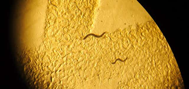
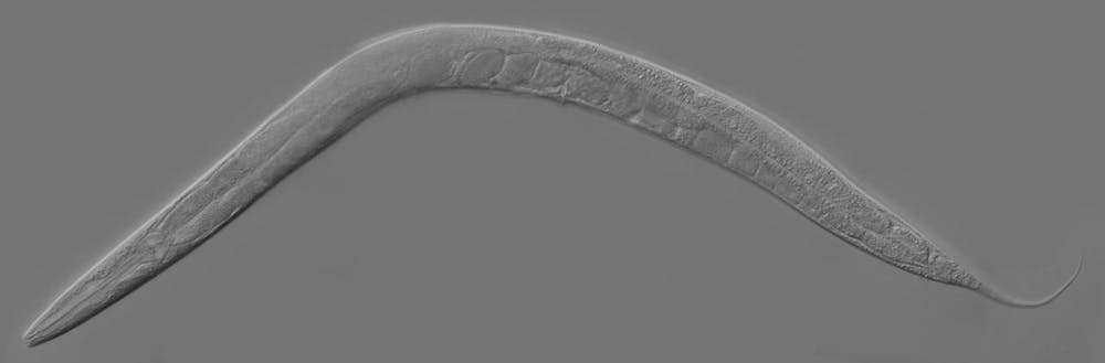
A small animal that could be easily cultivated and that had a short life cycle would be required. Ultimately, Brenner alighted on a nematode (roundworm) called Caenorhabditis elegans.
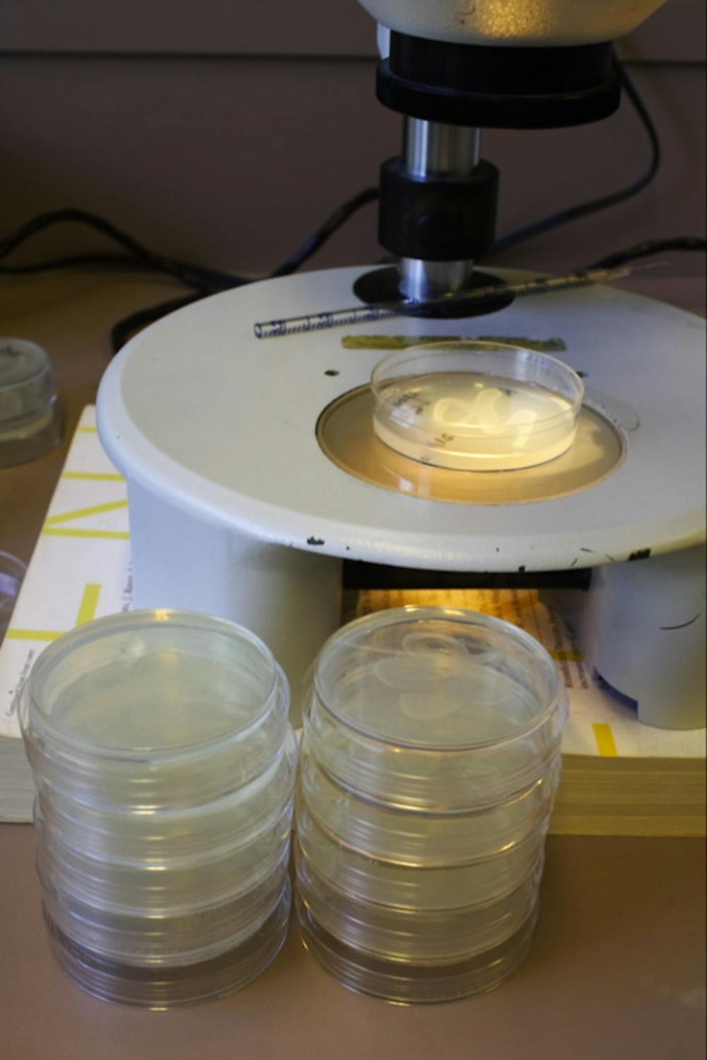
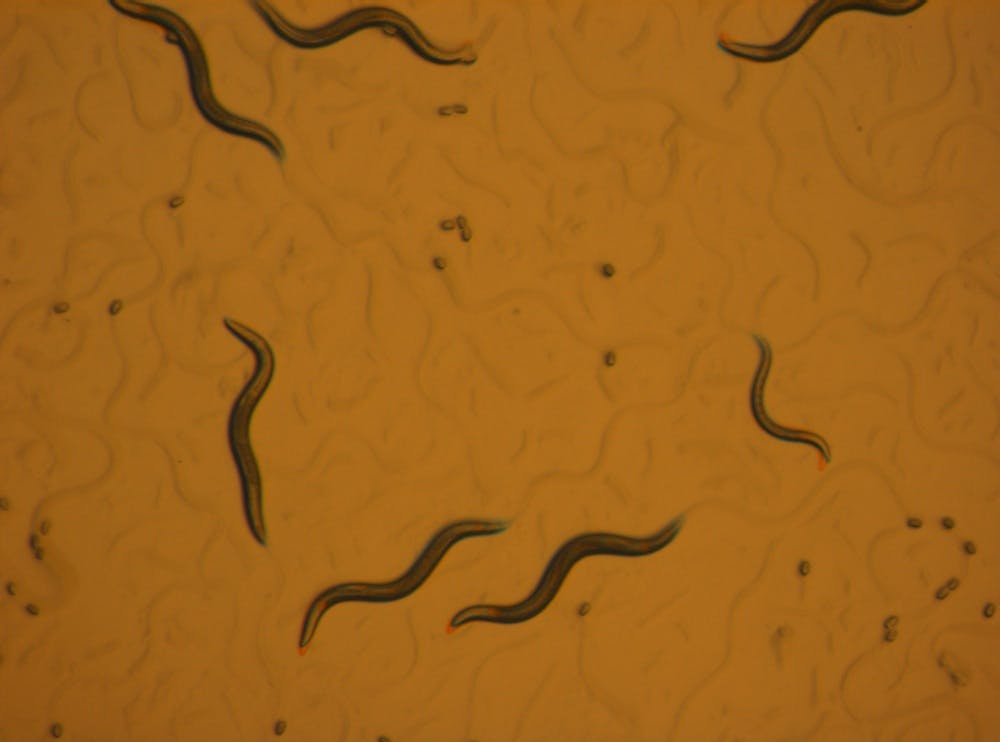
Measuring only 1mm, growing readily on simple agar plates with bacteria for food, and taking only three days to develop from an egg to a fertile hermaphrodite adult, the nematode fitted the bill.
More commonly referred to simply as “the worm”, Brenner’s model organism of choice is now the subject of research in hundreds of laboratories around the globe, including, at my count, at least ten labs in Australia.
In each of these worm labs, researchers study animals derived from a single sample that was taken from mushroom compost in Bristol in the UK in the 1950s.
The elegant worm
From these humble beginnings, the worm has afforded us notable insights in many areas of biology.
In line with Brenner’s aspirations, C. elegans has proved to be an excellent tool for studying development.
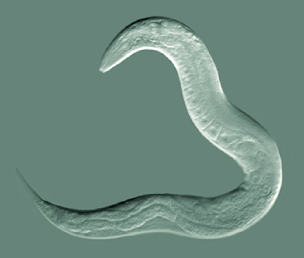
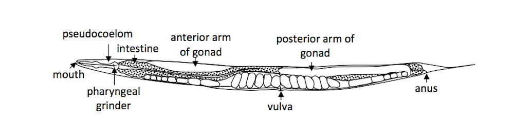
Remarkably, every worm grows in essentially the same way – from a single cell, the fertilised egg, to an adult, with precisely 959 somatic cells.
The worm is transparent, so these cells can be observed using light microscopy.
Through painstaking work requiring several years at the microscope, British biologist John Sulston meticulously mapped the process of worm development, documenting the timing and direction of every cell division.
This cell lineage is a vital tool that enables researchers to answer questions about how development is controlled at the level of a single cell.
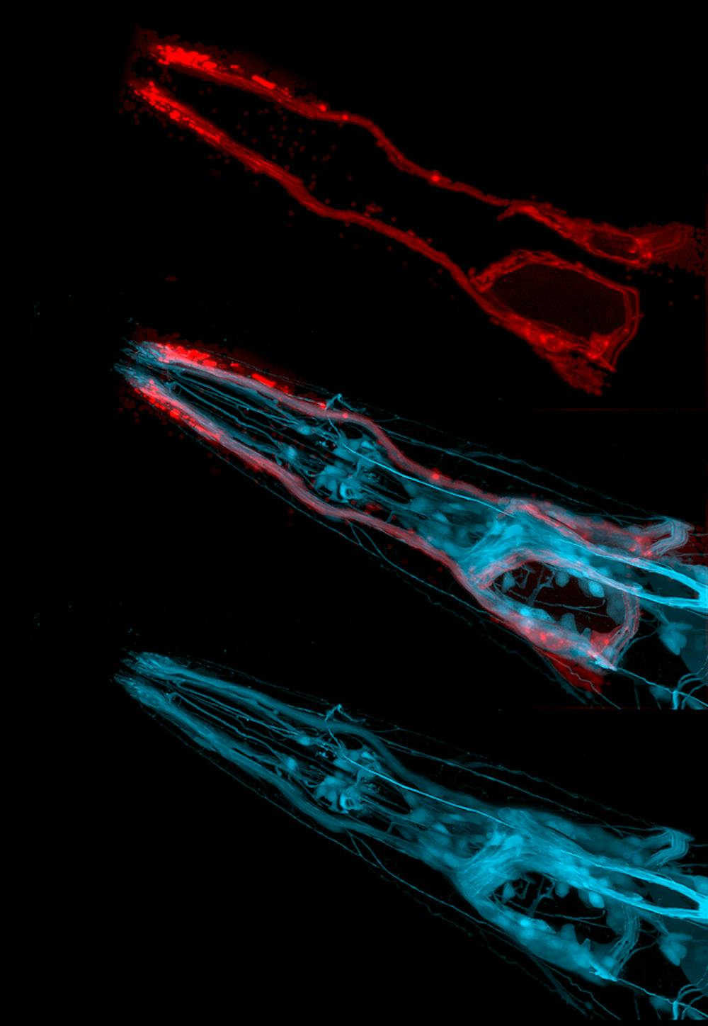
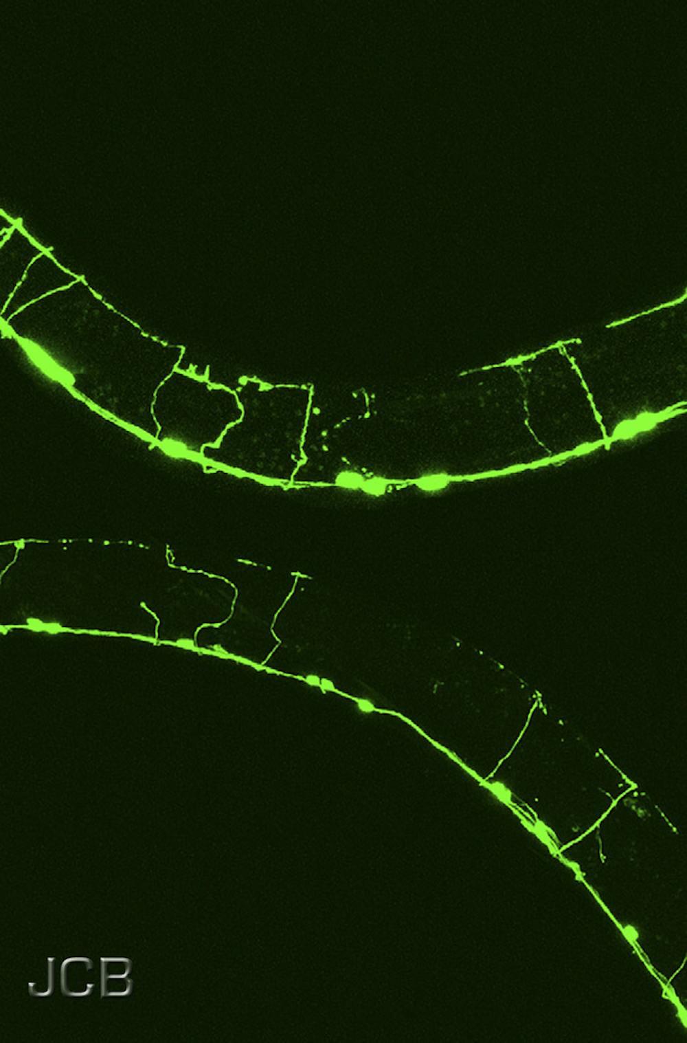
The worm has also lived up to the hope of providing insight into the workings of the nervous system.
Unlike our nervous system, which contains many billions of neurons, the worm has only 302 neurons.
Neurons are complex cells, which extend long processes and communicate between themselves, and with other cell types, via synapses.
After sectioning the worm into about 20,000 slices, American biologist John White and colleagues used electron microscopy to build a picture of all worm neurons, their processes and their 8,000 synapses, providing us with a complete wiring diagram of this relatively simply nervous system.
As with the developmental cell lineage, this neuronal wiring diagram is an important tool in enabling researchers to address many interesting questions relating to neuronal development and function.
Different strains
Despite the worm’s simplicity and the many obvious differences between worms and humans, if you look at what is going on inside our cells, we in fact have much in common. Take, for example, programmed cell death – the death of some of an organism’s cells as part of its natural development.
This process is vital for normal development but also plays an important role in cancer.
Seminal discoveries about programmed cell death were made using C. elegans as a model system and earned Brenner, Sulston and Robert Horvitz the Nobel Prize in Physiology or Medicine in 2002.
Another major discovery made using C. elegans was that of RNA interference, gene silencing by double-stranded RNA. For this, Americans Craig Mello and Andrew Fire were awarded the Nobel Prize in Physiology or Medicine in 2006.
Then, in 2008, American worm researcher Martin Chalfie shared in the Nobel Prize in Chemistry for his contribution to the development of green fluorescent protein as a tool for visualising biological structures in living organisms.
Aside from these seminal Nobel-winning discoveries, research using C. elegans continues to yield new information in diverse areas of biology such as behaviour, metabolism and ageing, to name but a few.
Many resources are shared among the worm research community, facilitating these endeavours.
These include an extensive collection of worm strains carrying mutations in particular genes and bacterial strains that can be fed to worms to reduce the function of any gene of interest by RNA interference.
Since around 40% of the genes implicated in human disease have equivalents in worms, these tools also make it possible to learn more about the underlying mechanisms of many diseases by examining the function of the related gene in the worm.
It is also straightforward to introduce new genes into C. elegans, including those from humans.
This approach has, for example, been used to create worm models for studying Alzheimer’s disease.
Such disease models can further be exploited to screen for drugs which may ultimately be used to treat human disease.
Through the past 50 years C. elegans has proved to be a very powerful model organism, revealing to us important lessons about our own biology.
The collaborative spirit that characterises the C. elegans research community, which is epitomised in the publication of The Worm Breeders Gazette, a newsletter for sharing results and techniques, will ensure that the next half century in worm world is equally productive.
To read more in the Animals in Research series, follow the links below:
Drosophila melanogaster (the fruit fly)
Danio rerio (zebrafish)