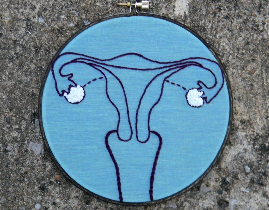
Ovarian cancer is the fifth highest killer in women when it comes to cancer, according to statistics from the American Cancer Society, with about 14,000 deaths in the United States alone this year. The disease is particularly nasty because it starts to spread to other tissues much earlier than other cancers do.
Once the spread starts, the tissue that suffers first tends to be belly fat. Previously it was thought this spreading occurred through simpler means. However, new research published in the journal Cancer Cell shows that the spreading actually happens via the blood. This could help develop new ways to prevent the spread.
The fatty tissue in the abdomen is a prime target for ovarian cancer cells because they produce a protein called NRG1. Once cancer cells have access to NRG1, they activate another protein, called HER3, which lets them multiply rapidly. Once the spread begins to the fat cells in the abdominal area, cancer cells can spread easily to the liver or lungs.
Since that abdominal area is so close to the ovary, researchers had assumed that the cancer cells simply detach from the ovary and float in the fluid filling the area till they reach the abdominal area. Yet, doctors would often observe that patients had tumours at the side of the fatty tissue that was quite far away from the ovary. Sometimes patients would show metastatic tumours in other organs like the liver or lungs but not in the abdominal area surrounding the ovary. Could it be that cancer cells were also using blood vessels to make their escape?
Researchers at MD Anderson Cancer Centre of the University of Texas, led by cancer biologist Anil Sood, came up with an innovative experiment to find the route cancer cells take. They physically joined two mice by stitching their skin together from hip to shoulder. After the surgery, the two mice ended up sharing a common blood circulatory system with their blood vessels fusing.
One of the mice among the artificially conjoined twins was made to develop ovarian cancer. If only the twin with ovarian cancer saw a spread of cancer to the abdominal area, it would prove that the previous theory about how ovarian cancer spread was right. But, instead, if the both twins saw development of cancer in their respective abdominal area, then the only way that could happen was through spreading via the blood.
Sood’s observation confirmed the latter scenario. Since the healthy twin’s abdomen was not physically close to the diseased ovary but still develop cancer, those cancer cells must have spread via the blood.
Interestingly, comparing the cancer cells that reached the healthy twin with the ones in the diseased ovary, the researchers found the travelling cancer cells were producing high amounts of a protein called HER3. In fact, cancer cells with more HER3 formed larger tumours in the abdominal area. And reducing the amount of HER3 protein in the cancer cells shrank the tumours in that area.
Sood thinks what preferentially brought the cancer cells to the healthy twin’s abdominal fat was the NRG1 protein, which then switches on HER3. To confirm that this happens in humans too, Sood checked patient records and found that those who had higher levels of HER3 protein in the tumour succumbed to the disease faster.
The discovery could help in developing better treatments. HER3 is a close cousin of the protein HER2 that is famous for being a drug target in breast cancer. Drugs that block HER2 may work against HER3 as well. In fact, a drug called pertuzumab, developed for breast cancer patients with high levels of HER2, is currently undergoing clinical trials for patients with ovarian cancer, conducted by Hoffmann-La Roche.
Next, read this: Now we know why drugs don’t work on pancreatic cancer

